Golgi Apparatus: What Is It, Location, Functions, and More
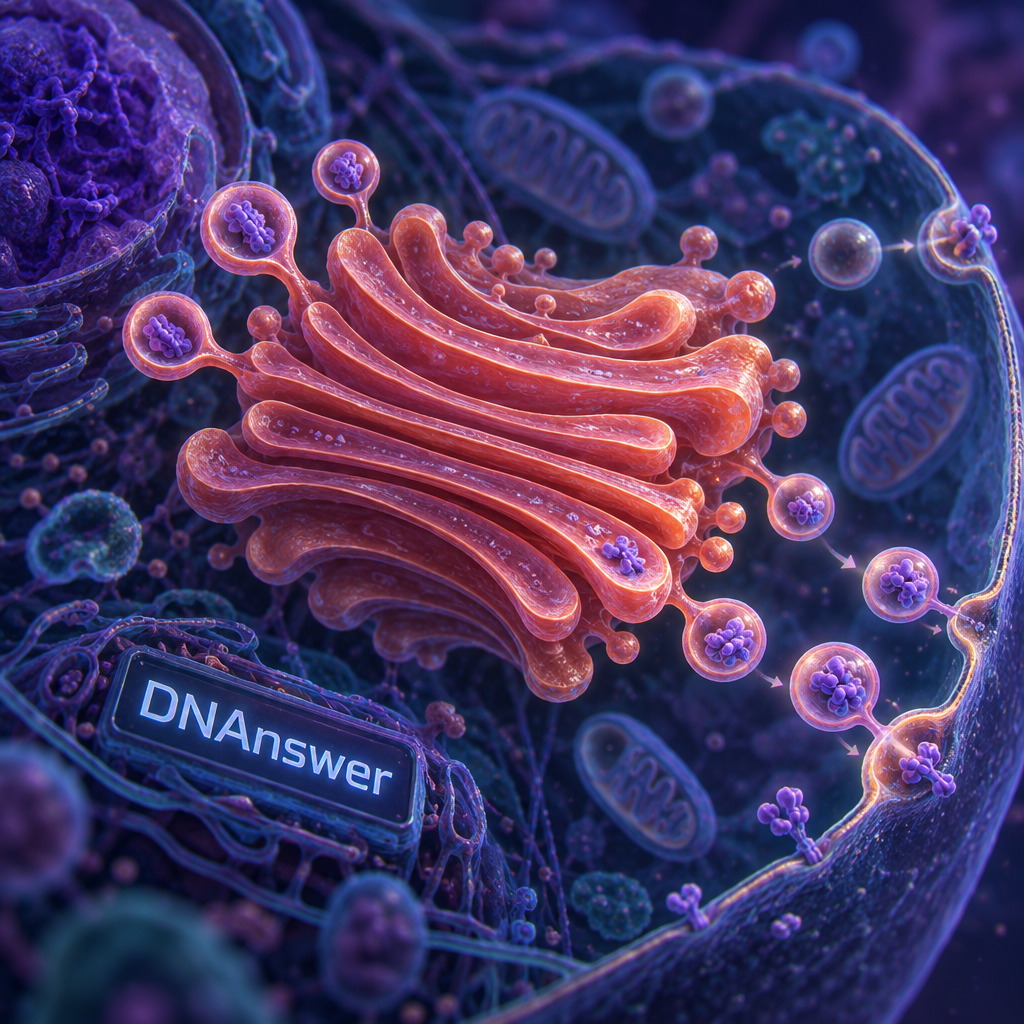
Is the Golgi apparatus really a cellular post office? That's the line most of us remember from biology class, and it's only a quarter true. The functions of the Golgi apparatus include editing, tagging, and sorting cargo, and the tags it adds decide where each protein goes. A post office never rewrites your mail. The Golgi does.
Key Takeaways
- The Golgi modifies proteins, not just ships them.
- Sugar tags added there act like built-in shipping addresses.
- It sits near the nucleus, fed by vesicles from the ER.
- Cargo moves cis to trans, being edited at each stop.
- Size and shape vary by cell type; bacteria have none.
- Golgi failure underlies several inherited diseases.
Myth 1: "The Golgi Is a Cellular Post Office"
The belief is that the Golgi receives packages and forwards them unchanged. Teachers use this picture for good reason. It captures one real thing: the Golgi receives cargo from one place and sends it to others. For a first pass at biology, that's a fair shortcut.
The trouble is what the analogy leaves out. A post office doesn't open your parcel, swap parts, and stick on a new label. The Golgi does exactly that, and the changes matter.
What actually happens inside
Proteins arrive from the endoplasmic reticulum (ER), the folded-membrane factory where proteins are first built. They enter the Golgi still unfinished. Enzymes, which are proteins that speed up chemical reactions, trim sugar chains, add new sugars, attach phosphate groups, and sometimes cut the protein itself into its active form.
Insulin shows the point. Your pancreatic cells make it as a longer precursor, and cutting steps along the secretory route (the path out of the cell) produce the working hormone. Without those edits, the molecule can't do its job.
Why the myth costs you
If you think the Golgi is passive, you'll misread questions about disease, drugs, and exams alike. Many inherited disorders come from broken editing, not broken delivery. A better picture is a customization factory with a sorting hub attached. I'd keep the post office image only for the last step: the shipping.
The Golgi's name comes from Camillo Golgi, the Italian scientist who described it in 1898 using a silver-based stain. Some scientists doubted for decades that it was real, suspecting the stain created it. Electron microscopes in the 1950s settled the question.
Myth 2: "The Golgi Just Packages and Ships Proteins"
The belief is that the Golgi's whole job is boxing things up. This one comes from the word "packaging" itself, which appears in textbooks beside the organelle (a small structure with a job inside the cell). Packaging is real. Cargo does leave the Golgi in membrane bubbles called vesicles.
But packaging comes last. Before it, cargo gets processed in a set order, stack by stack.
The assembly line: cis, medial, trans
The Golgi is a pile of flattened sacs called cisternae (singular: cisterna). Think of a stack of deflated pita breads. The side facing the ER is the cis face, the middle is medial, and the side facing away is the trans face. Different enzymes live in different sacs.
Here is the route cargo follows:
- Vesicles from the ER fuse with the cis face.
- Early enzymes trim sugar chains on the cargo.
- In the medial sacs, new sugars attach.
- Enzymes add sulfate or phosphate groups where needed.
- At the trans face, the cargo gets sorted by its tags.
- Vesicles bud off toward their destinations.
What this means for you
Packaging is the finale of a multi-step edit. When a quiz asks what the Golgi does, "modifies, sorts, and packages" is the full answer, in that order. Modification comes first, and without it, the sorting has nothing to read.
One honest caveat: scientists still debate exactly how cargo moves through the stack. One model says the sacs themselves mature and shift from cis to trans. Another says vesicles carry cargo between fixed sacs. Evidence supports parts of both, and the question remains open.
Myth 3: "Sugar Tags Are Just Decoration"
Sugar sounds like a coating, so people assume it's cosmetic. That's an understandable guess. Sugars on a protein look like icing on a cake.
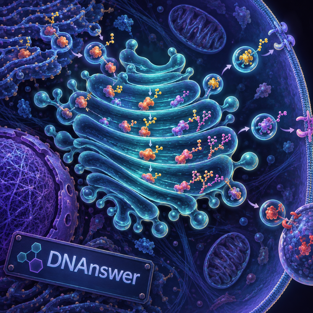
They work more like a barcode. Glycosylation (adding sugar chains to a protein) changes how a protein folds, how long it survives, and where it goes. Cells don't add these chains by accident. Enzymes called glycosyltransferases build each one in a controlled sequence.
Tags that act as addresses
The clearest example is the lysosome, the cell's recycling bin. Enzymes headed there get a sugar tagged with phosphate, called mannose-6-phosphate. A receptor in the trans Golgi recognizes that tag and diverts those proteins into vesicles bound for lysosomes. No tag, no trip.
Your blood type also depends on sugar tags. The A and B antigens, which doctors check before a transfusion, are sugar structures added to proteins and fats on red blood cells. Different sugar patterns, different blood types.
What to do with this
Remember that sugar tags are the address system. When you meet a disease caused by missing enzymes, ask which tag failed. That question usually points to the answer faster than memorizing the disease name.
I-cell disease is caused by failure to add the mannose-6-phosphate tag, so lysosomal enzymes get secreted from the cell instead of reaching the lysosome.
Doctors call that condition I-cell disease (mucolipidosis II), and it shows how much one missing tag can matter.
Myth 4: "The Golgi and the ER Work Alone"
The belief is that each organelle has its own job and runs independently. School diagrams encourage this, since each structure gets its own neat label and color.
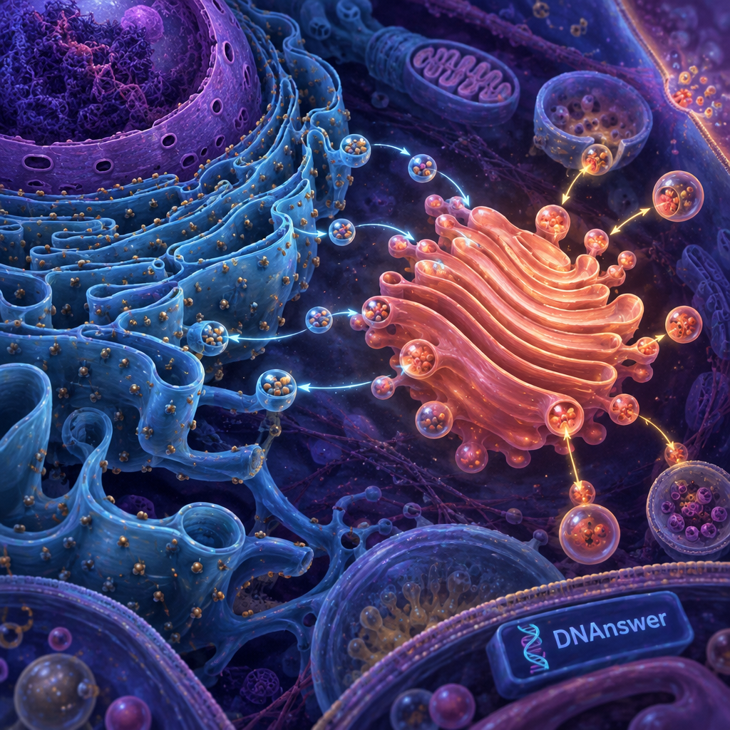
In a living cell, the ER and Golgi form one pipeline. The ER builds proteins and folds them. Then coated vesicles carry them across a short gap to the Golgi. A protein coat called COPII wraps vesicles leaving the ER, and COPI wraps vesicles returning cargo the other way.
Traffic goes both directions
Return traffic matters because the Golgi must send escaped ER proteins back home. Many ER residents carry a short amino acid signal, KDEL, that gets them recycled. Without that system, the ER would slowly lose its own equipment.
Where the Golgi sits
In animal cells, the Golgi usually sits close to the nucleus, the compartment holding your DNA, and near the ER. Tubes of the cell's internal scaffolding, called microtubules, hold it there. Break those tubes with a drug, and the Golgi fragments and scatters around the cell.
So when you picture the organelle, put it in a neighborhood. Location tells you its job: it sits at the end of the ER's production line. If you're studying for a test, draw the two together with arrows between them.
Myth 5: "Every Cell Has the Same Golgi"
The belief is that the Golgi is a fixed stack of about six sacs in every cell. Diagrams reinforce it, because a textbook has room to draw only one tidy version.
Real Golgi structures vary a lot. A cell that secretes heavily, like a goblet cell lining your airways that makes mucus, has an enlarged Golgi. A resting cell has a smaller one. Some cells hold hundreds of small stacks instead of one large one.
Plants, animals, and bacteria
Plant cells often have many separate stacks scattered through the cytoplasm, the jelly-like interior. Animal cells usually link their stacks into a single ribbon near the nucleus. Plant Golgi stacks also build cell wall material, including pectin and hemicellulose, which are sugar-based fibers that give plant tissue its firmness.
Bacteria have no Golgi at all. They're prokaryotes, meaning their cells have no membrane-bound organelles. Archaea lack one too. Only eukaryotes, the group including animals, plants, and fungi, have this structure.
Practical takeaway
Treat any diagram as a sketch. If a question asks about a specific cell, think about what that cell makes. A plant cell building walls and an antibody-secreting immune cell both lean on the Golgi, but they use it differently.
Myth 6: "The Golgi Only Handles Proteins"
People believe this because most lessons follow proteins. It's an easy storyline, so it dominates.
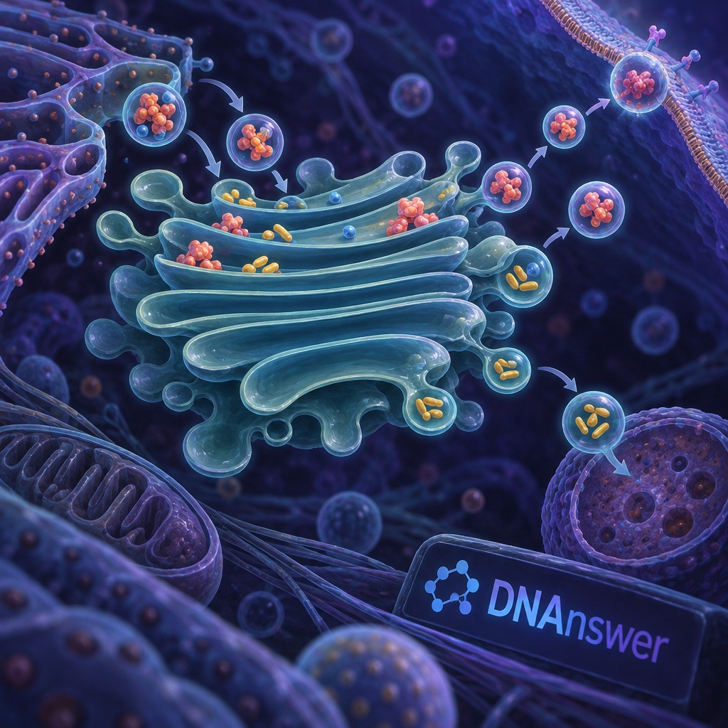
The Golgi also handles fats. It helps build sphingomyelin, a lipid (fat-type molecule) in cell membranes, and it makes glycolipids, which are fats with sugar chains attached. It also sorts lipids along with proteins into the right membranes.
Membrane is cargo too
Each vesicle that leaves the Golgi carries a patch of membrane. When a vesicle fuses with the cell surface during exocytosis (release of contents outside the cell), that membrane merges into the outer boundary. The cell's surface constantly gets renewed this way.
Where cargo ends up
Vesicles from the trans face have three main destinations. Some head to the cell surface and release hormones, enzymes, or mucus. Others form lysosomes. Still others replace membrane or deliver proteins to the surface permanently. Think of the trans Golgi network as the sorting hub, reading tags and routing cargo to one of these three exits.
When the Golgi Fails: Disease Connections
You might expect a broken Golgi to be rare and strange. Several conditions trace back to it. Congenital disorders of glycosylation (CDG) are a family of inherited conditions where sugar-tagging enzymes don't work, and they can affect the brain, liver, and muscles.
Researchers have also reported Golgi fragmentation in neurons from people with Alzheimer's disease and ALS, though scientists still debate whether it causes damage or follows it. That's a drawback of drawing neat conclusions here: the link is clear, the direction isn't.
If you want to test your grip on organelles like this one, the quizzes at dnanswer.app work well for practice.
Conclusion
Next time you see "post office," upgrade it. Picture a factory that edits each item, stamps an address in sugar, then dispatches it. Use that picture on tests and when reading health news. Choose "modifies, sorts, and ships" as your default answer, and ask which tag or enzyme is involved whenever a disease touches protein delivery.
Frequently Asked Questions
Where is the Golgi apparatus located in a cell?
In animal cells, it sits near the nucleus and next to the ER. Plant cells often scatter many smaller stacks through the cytoplasm. Microtubules help anchor the animal Golgi in place.
What is the main function of the Golgi apparatus?
Its main job is to modify, sort, and package proteins and lipids from the ER. Glycosylation, the addition of sugar chains, is the best-known modification, and those sugars guide each molecule to its destination.
Do all cells have a Golgi apparatus?
No. Bacteria and archaea lack it. Nearly all eukaryotic cells have one, though mature red blood cells in mammals lose their organelles during development.
Who discovered the Golgi apparatus?
Camillo Golgi, an Italian physician, described it in 1898 after staining nerve cells with silver. He shared the 1906 Nobel Prize in Physiology or Medicine with Santiago Ramón y Cajal for work on the nervous system.