Endoplasmic reticulum
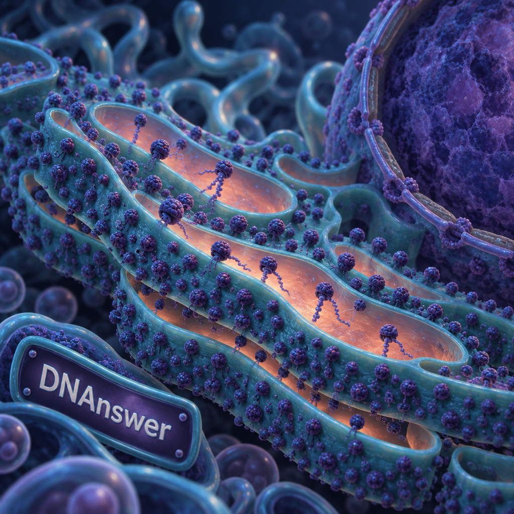
Is the bumpy ER really a separate protein factory with ribosomes built into its walls? No. The rough ER is a region of one continuous membrane, and its "bumps" are ribosomes that dock only when a protein's first few amino acids demand it.
If you remember the textbook line "rough ER makes proteins," you have about 30% of the story. This article corrects five common beliefs about the endoplasmic reticulum rough region, and shows why the corrections matter for diseases like cystic fibrosis.
Key Takeaways
- Rough and smooth ER are one connected membrane, not two organelles.
- Ribosomes dock temporarily; a signal peptide decides which ones.
- Free ribosomes make most proteins that stay inside the cell fluid.
- The rough ER inspects proteins and rejects faulty ones.
- Too many misfolded proteins trigger the unfolded protein response.
- Misfolding drives cystic fibrosis and other diseases.
Myth 1: Rough and Smooth ER Are Two Separate Organelles
Open almost any textbook diagram and you see two neat objects: a stack of studded sheets and a tangle of smooth tubes. That picture is wrong. They are one membrane, with different neighborhoods.
The diagrams exist for a good reason. Under an electron microscope, a slice through a cell shows studded flat sheets in one spot and smooth tubes in another. Early microscopists named what they saw, and the names stuck. A snapshot makes parts look like separate things.
One membrane, one inner space
The ER is a single network of flattened sacs called cisternae (flat, fluid-filled pouches) and tubes. All of it encloses one continuous inner space, the lumen. A molecule that enters the lumen at a rough patch can travel to a smooth patch without ever crossing another membrane.
The network also connects to the nucleus. The outer layer of the nuclear envelope, the double membrane wrapped around your DNA, flows directly into the rough ER. The ER is, in effect, an extension of the nuclear membrane.
Regions change with the cell's needs
The "rough" and "smooth" labels describe jobs, not fixed borders. Studded sheets handle proteins headed for secretion or membranes. Smooth tubes handle fats and steroid hormones, and they store calcium. The balance shifts as needs shift.
Liver cells show this well. Give the body a drug such as phenobarbital, and liver cells expand their smooth ER to process it. Remove the drug and the smooth region shrinks again. Fixed organelles don't do that.
What should you do with this? Picture the ER as a landscape of one connected sheet, like a single crumpled bedsheet with some patches covered in sand. The sand can be added or removed.
Myth 2: The Bumps Are Part of the ER Itself
The bumpy surface looks like texture built into the membrane. It isn't. Each bump is a ribosome, a molecular machine that reads messenger RNA (mRNA, the working copy of a gene) and strings amino acids into a protein.
People believe the myth because the studding looks permanent in photographs. A frozen image can't show that ribosomes come and go. Early researchers, including George Palade, who shared the 1974 Nobel Prize in Physiology or Medicine for his work on cell structure, described ribosomes attached to the membrane. The attachment was real. The permanence was an assumption.
Ribosomes are separate machines. They are made of RNA and proteins, they float in the cell fluid (the cytosol), and they cycle on and off the ER. When a ribosome finishes a protein, it detaches and can start again anywhere.
Think of ribosomes as food trucks, not as bricks in the restaurant wall. A truck parks where an order demands it, then drives away. The ER membrane is the parking lot. It has docking sites, but the trucks arrive on demand.
Your takeaway: when you see "rough" in a diagram, read it as "ribosomes are visiting right now." The state is temporary and depends on what the cell is building.
Myth 3: Ribosomes Make Proteins Only on the ER
Many people leave biology class believing there are two kinds of ribosome, free ones and ER ones. The truth is stranger. There is only one kind. Every ribosome starts out free, and a single protein's instructions decide where it ends up.
How a ribosome picks its location
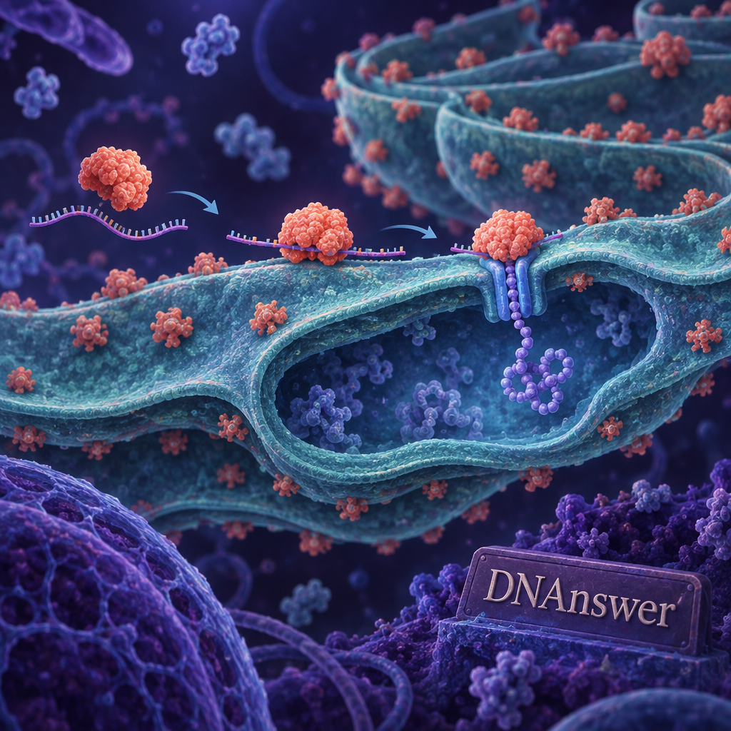
Every ribosome in a human cell starts translating mRNA in the cytosol. If the protein is meant to stay in the cytosol, such as the enzymes that break down sugar, the ribosome finishes the job right there. No ER involved.
If the protein is headed for secretion or for a membrane, its first 15 to 30 amino acids form a signal peptide, a short address tag. As that tag emerges from the ribosome, a particle called the signal recognition particle (SRP) grabs it. The SRP pauses translation and escorts the whole ribosome to the ER.
The docking sequence
Here is the order of events, from tag to threading.
- A ribosome starts reading an mRNA in the cytosol.
- The first amino acids emerge, forming a signal peptide.
- The SRP binds that signal and pauses protein building.
- The SRP carries the ribosome to an SRP receptor on the ER membrane.
- The ribosome locks onto a channel called the translocon (a protein pore through the membrane).
- The SRP lets go, and building resumes, with the growing chain pushed through the channel into the ER lumen.
This is co-translational import: the protein enters the ER while it is still being built. The cell doesn't finish a protein and then haul it in. It threads the chain through the pore as it grows.
Your takeaway: the "rough" look in a cell comes from mRNAs for secreted and membrane proteins. A cell making mostly internal enzymes would have sparse studding, even with plenty of ribosomes.
Myth 4: The Rough ER Makes All the Cell's Proteins
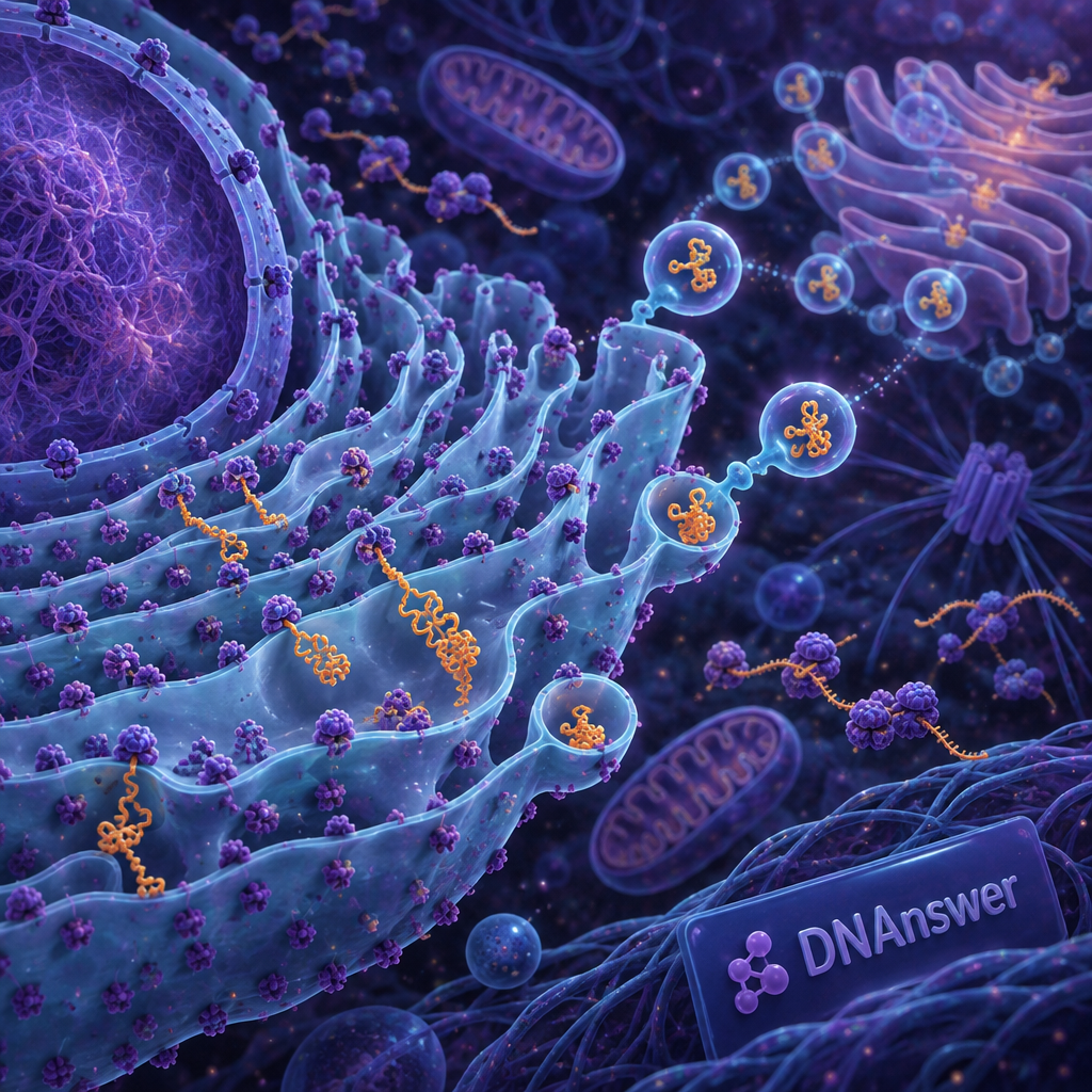
If rough ER is the protein factory, the cell's whole protein output must come through it. That idea is common and mostly an overreach of a true statement. The rough ER makes one important class of proteins, not all of them.
Three groups of proteins get made on rough ER ribosomes. The first is secreted proteins, such as insulin and antibodies. The second is membrane proteins, including the receptors on your cell's surface. The third is residents of the compartments along the route, such as proteins that live in the Golgi or in lysosomes (the cell's recycling bins).
Everything else is made by free ribosomes. That includes the proteins that fold DNA, build the cell's internal scaffold, and run basic metabolism. Some proteins go to the nucleus, mitochondria, or peroxisomes with their own separate tags and routes.
Rough estimates suggest that roughly a third of human proteins are processed through the ER route. Treat that as an approximation, since counts vary with the method used and with the cell type. The ratio shifts widely from one tissue to another.
Who has the most rough ER?
Cells that export huge amounts of protein build the most rough ER. Plasma cells, the immune cells that make antibodies, can pump out thousands of antibody molecules per second, and their cytoplasm fills with studded sheets. Pancreatic acinar cells, which make digestive enzymes, look much the same. So do the pancreatic beta cells that make insulin.
A muscle cell, by contrast, makes mostly internal proteins and has far less rough ER. Different jobs, different architecture.
What to do with this: when you read that a cell is "full of rough ER," translate it to "this cell ships a lot of protein out or into its membrane." The studding tells you about the cell's job.
Myth 5: The ER Is Just a Static Protein Factory
This is the myth that costs the most, because it hides the ER's real work: inspection. A factory implies a line that makes products and ships them. The rough ER behaves more like a factory with a strict quality-control department, and the department can shut down production.
The factory picture has a kernel of truth. Proteins do enter the ER as raw chains and leave as finished products. But entering is only the first step, and the cell scrutinizes what comes through.
Folding, sugar tags and chaperones
Once a chain enters the lumen, it must fold into a precise 3D shape. Helper proteins called chaperones (proteins that guide folding and stop sticky clumps) assist this. Enzymes also form disulfide bonds, which are sulfur-to-sulfur links that lock a shape in place.
Another step is N-linked glycosylation: the cell attaches a preassembled sugar cluster to specific asparagine amino acids. These sugars act as a status label. Enzymes trim the sugar as the protein folds, and a protein that stays misfolded gets re-tagged for another try or for disposal.
Rejection and the stress response
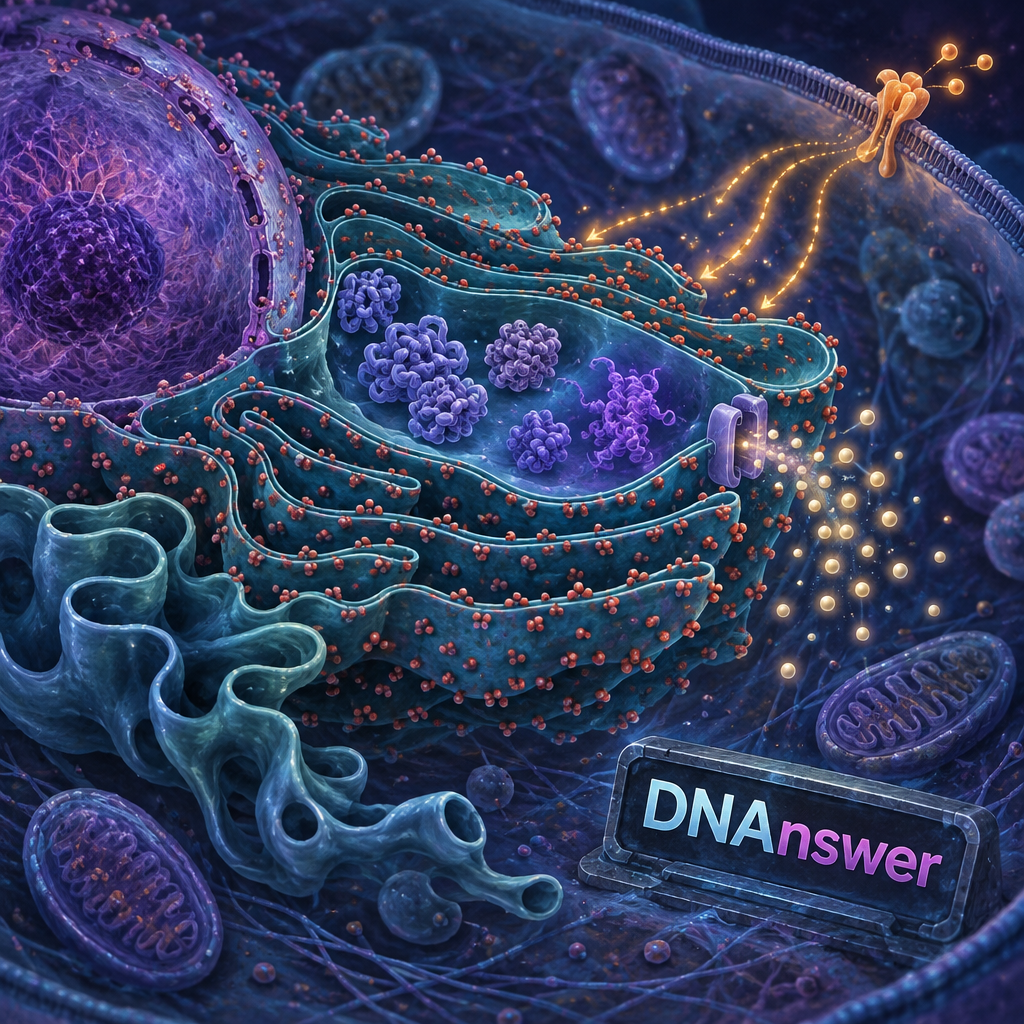
Proteins that fail inspection get pulled back out through a channel and destroyed in the cytosol, a process called ER-associated degradation. If misfolded proteins pile up faster than the ER can clear them, the cell sounds an alarm known as the unfolded protein response.
The response does three things. It slows new protein production, boosts chaperone supply, and expands the ER. If stress never eases, the same pathway can switch to a cell-death program. The cell sacrifices itself rather than ship bad product.
The unfolded protein response is a signaling network that detects ER stress and adjusts the ER's capacity to match its workload.
That definition fits how the field describes it, and it is worth keeping: the ER measures its own workload.
Your takeaway: stop picturing a conveyor belt. Picture a checkpoint that can halt traffic.
When Quality Control Fails: Real Diseases
The inspection system matters most when it goes wrong. Many inherited and age-related diseases trace back to proteins that misfold in the ER.
Cystic fibrosis is the classic example. The CFTR gene makes a channel that moves chloride across cell membranes. The most common mutation, called F508del, removes one amino acid. The protein would still work if it reached the surface, but it misfolds slightly, and the ER's quality control rejects it. Without the channel, mucus in the lungs turns thick and sticky.
That discovery changed treatment. Drugs known as CFTR modulators help the mutant protein fold or work better. One example is the combination therapy Trikafta, approved by the U.S. Food and Drug Administration in October 2019 for many people with cystic fibrosis. The drug doesn't replace the gene. It helps the faulty protein pass or function.
Other conditions link to ER stress as well. Alpha-1 antitrypsin deficiency involves a misfolded protein that clogs liver cells. Some forms of diabetes involve stressed insulin-making cells. Researchers also study ER stress in neurodegenerative diseases, though the exact role there is still debated.
One drawback of this framing: ER stress is a piece of the puzzle in many diseases, not the single cause. Be wary of headlines that blame it for everything.
If you want to test your grasp of organelles like this one, the quizzes at dnanswer.app are a low-stakes way to check what stuck.
Conclusion
Next time you see a diagram of the endoplasmic reticulum rough region, redraw it mentally. One continuous membrane. Ribosomes docking only when a signal peptide calls them. A checkpoint instead of a conveyor belt. That shift helps when you read about drugs like CFTR modulators, because the treatment target is often the folding step. When a headline says "protein factory," ask what gets inspected, and what gets rejected.
Frequently Asked Questions
Why is it called rough endoplasmic reticulum?
It looks rough under an electron microscope because ribosomes dot its outer surface. Endoplasmic means inside the cell fluid, and reticulum means network. Cells with little ribosome docking show a smoother appearance in those regions.
What is the difference between rough and smooth ER?
Rough ER carries ribosomes and handles proteins for secretion and membranes. Smooth ER has none and makes lipids, processes drugs and toxins in the liver, and stores calcium. They connect as one membrane, and a cell can reshape the balance.
Do plant cells have rough ER?
Yes. Plant cells have rough ER, and it connects to the nuclear envelope just as in animal cells. It builds secreted and membrane proteins, including those that help construct the cell wall.
Can a cell survive without a working ER?
No. The ER makes membrane proteins and handles calcium, so cells can't live long without it. Severe, unresolved ER stress triggers the cell to die by programmed cell death rather than keep running.