Cell Membrane (Plasma Membrane): Structure and Function
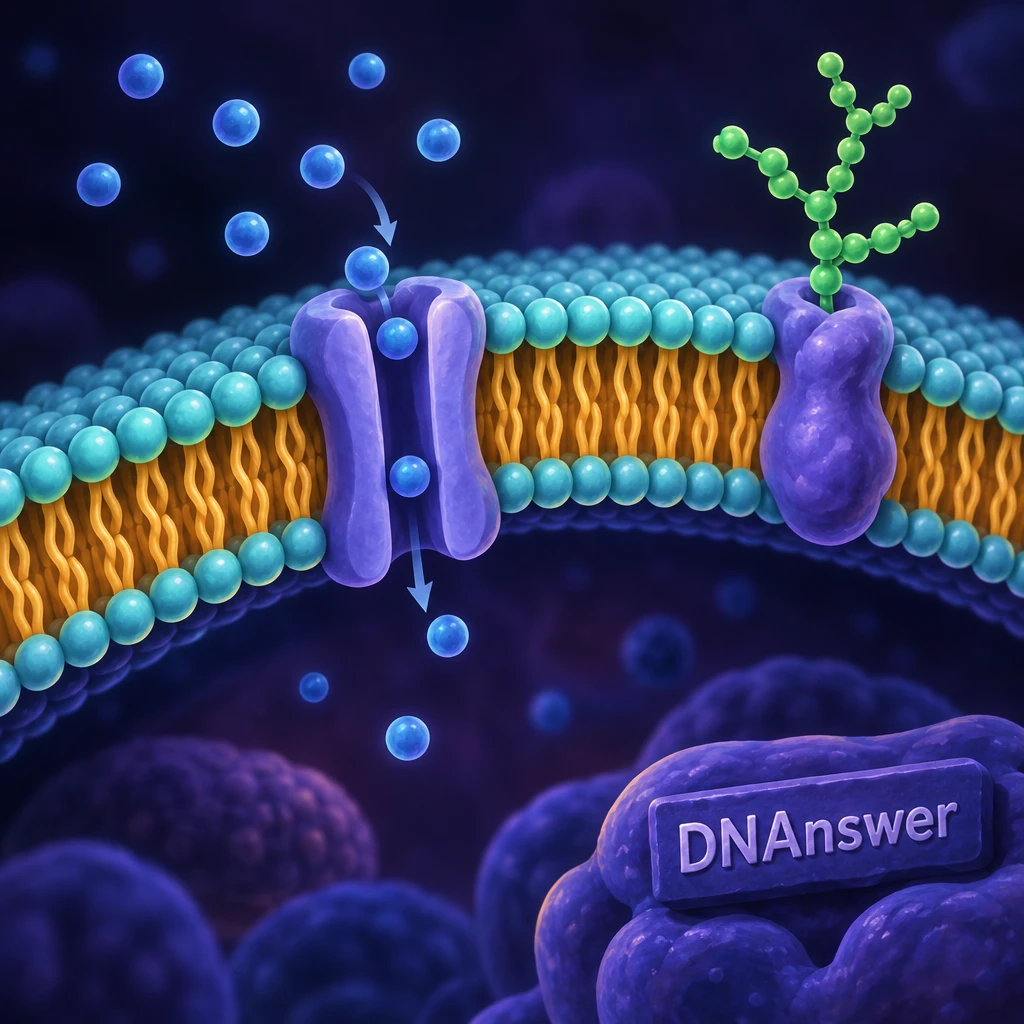
Is the cell membrane just a thin wall that keeps the cell's insides from spilling out? Not quite. It behaves more like a film of oil, about as runny as salad dressing, and it still decides what enters, what leaves, and which messages get through.
The cell membrane (biologists also call it the plasma membrane) wraps every living cell on Earth. Most of what you remember from school diagrams makes it look stiff, simple and passive. The real thing moves, sorts molecules by their chemistry, and talks to its neighbors. Clearing up the myths gives you a far more accurate picture of how your own cells stay alive.
Key Takeaways
- Cell membrane and plasma membrane mean the same structure.
- About 4 nanometers thick, and its parts move constantly.
- Every cell has a membrane; plants and bacteria add walls.
- Size alone doesn't decide what crosses. Charge and proteins matter too.
- Cholesterol keeps animal membranes from getting too stiff or runny.
- Membranes also handle recognition, signaling and attachment.
Myth 1: The membrane is a solid, fixed wall
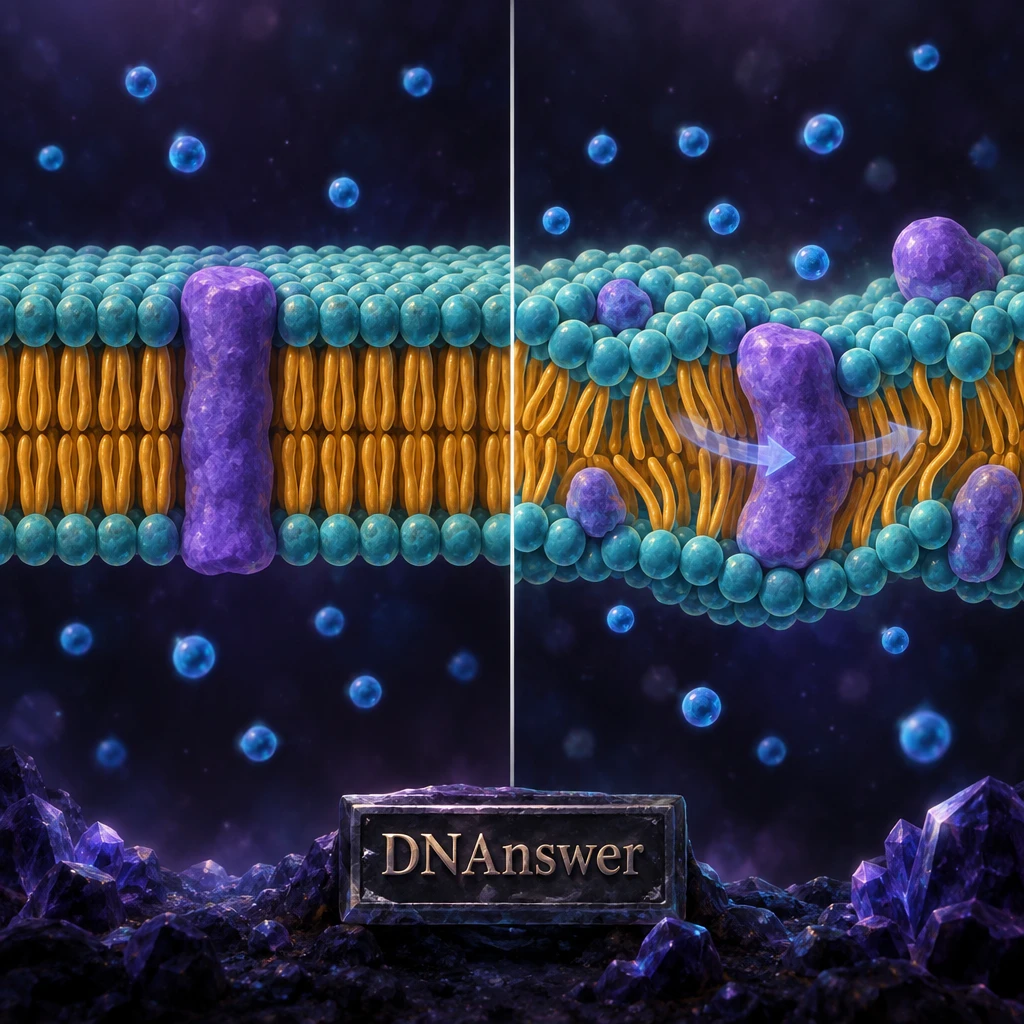
Picture the classic textbook drawing. Two neat rows of lollipop shapes, a few blobs sitting in them, everything frozen in place. No wonder most people imagine a hard shell.
That drawing has a fair reason to exist. Diagrams need to stand still, and the membrane really does keep the cell's contents separate from the outside. But a diagram works like one frame from a video. Scientists Seymour Singer and Garth Nicolson proposed the fluid mosaic model in 1972, and it still forms the basis of how biologists describe membranes today.
What the membrane actually looks like
The main building block is the phospholipid, a fat-like molecule with a head that likes water and two tails that avoid it. Put millions of them in water and they arrange themselves into two layers, tails tucked inward, heads facing the watery inside and outside of the cell. We call this a phospholipid bilayer.
Into that oily sheet the cell inserts proteins, cholesterol and sugar chains. By mass, a typical human cell membrane contains about 50 percent protein, 40 percent lipid (fats of all kinds) and 10 percent carbohydrate. That mix of different pieces explains the word "mosaic." The pieces slide past each other constantly, which explains "fluid."
Fluid mosaic model: the plasma membrane as a moving mosaic of phospholipids, cholesterol and proteins that drift freely within the plane of the membrane.
Thin, but not flimsy
How thin? A single phospholipid measures about 2 nanometers from head to tail, so the bilayer comes to roughly 4 nanometers. A nanometer equals one billionth of a meter. You could stack about 25,000 membranes to match the thickness of one sheet of printer paper.
Researchers who study cell sizes compare the proportions to an airplane. A plane's skin runs about 1 cm thick around a body about 5 m wide, a ratio of 1 to 500. A 4-nanometer membrane around a cell 2 microns wide gives the same 1 to 500 ratio.
The fluidity has a striking consequence. Push an extremely fine needle into a cell and the membrane flows around it. Pull the needle out and the membrane seals itself again. A rigid wall would crack instead. So when you picture a cell, swap the brick wall for a soap bubble that knows its job.
Myth 2: Plant and bacterial cells have a wall instead of a membrane
You probably learned that plant cells have cell walls and animal cells don't. Many people then assume the wall replaces the membrane in plants. It sounds logical: why would a cell need two boundaries?
The kernel of truth: cell walls really are tougher. Plants, bacteria, algae and fungi build a rigid outer layer, and it gives them far more structural strength than a plasma membrane ever could. That wall lets a tree stand upright and keeps a bacterium from bursting.
But every cell has a plasma membrane, with no exceptions. In plants and bacteria, the wall sits outside the membrane, like a cardboard box around a balloon. The wall gives shape and protection. The membrane still does the choosing: which nutrients enter, which waste leaves, which signals get read.
The wall can't do that job because it works more like a coarse mesh. Water and many small molecules pass through it easily. The fine control happens one layer in, at the membrane.
This matters when you read about antibiotics. Penicillin attacks the bacterial cell wall, which your own cells lack, so it harms bacteria far more than it harms you. Other drugs target membranes directly, and those behave very differently. When you see "cell wall" and "cell membrane" in the same sentence, read them as two separate structures doing two separate jobs.
Myth 3: Small things slip through, big things get blocked
Here's the version most of us carry around. The membrane works like a sieve, so tiny molecules pass and large ones don't. Size certainly plays a part, and that kernel of truth makes the myth stick.
Now test it. A water molecule is tiny, yet it crosses the bare lipid layer only slowly. A sodium ion (a charged sodium atom, the "sodium" in table salt) is tinier still, and the lipid layer blocks it almost completely. Meanwhile oxygen and carbon dioxide slip straight through. Size alone can't explain that pattern.
The four things that decide
Selective permeability means the membrane lets some substances cross and stops others. Four features decide the outcome:
- Size. Smaller molecules generally cross more easily.
- Charge. Charged particles, called ions, get repelled by the oily core.
- Chemistry. Fat-friendly molecules dissolve into the lipid layer. Water-loving ones struggle.
- Available proteins. If the right channel or carrier exists, almost anything can get through.
The charge rule surprises people most. Water molecules cling to an ion, wrapping it in a shell. To enter the oily core, the ion would have to shed that shell, and that costs a lot of energy. Researchers have measured this directly: the more water a molecule holds onto, the harder it finds the crossing.
Following familiar molecules
Trace a few molecules you already know. Oxygen, small and uncharged, crosses freely. Glucose, the sugar your cells burn for fuel, is too large and too water-loving to cross the lipid layer alone, so your cells use dedicated carrier proteins. Potassium and sodium ions need channels or pumps. Steroid hormones such as estrogen and testosterone, built from a fat-like skeleton, dissolve right through and act inside the cell.
So the better mental image looks less like a sieve and more like a building with a fat-soluble side entrance and many staffed doors. Who gets in depends on the visitor's chemistry and on which doors the cell has opened.
Myth 4: Passive transport just means leaking through the membrane
This one comes straight from simplified explanations. "Passive" sounds like "nothing happens," so people picture molecules drifting through the lipid layer on their own, while anything involving proteins must be active work.
The origin makes sense. Simple diffusion (molecules spreading from a crowded area to an emptier one, straight through the lipids) gives the cleanest example of passive transport. Textbooks show it first, and it sticks.
Passive transport can use proteins too
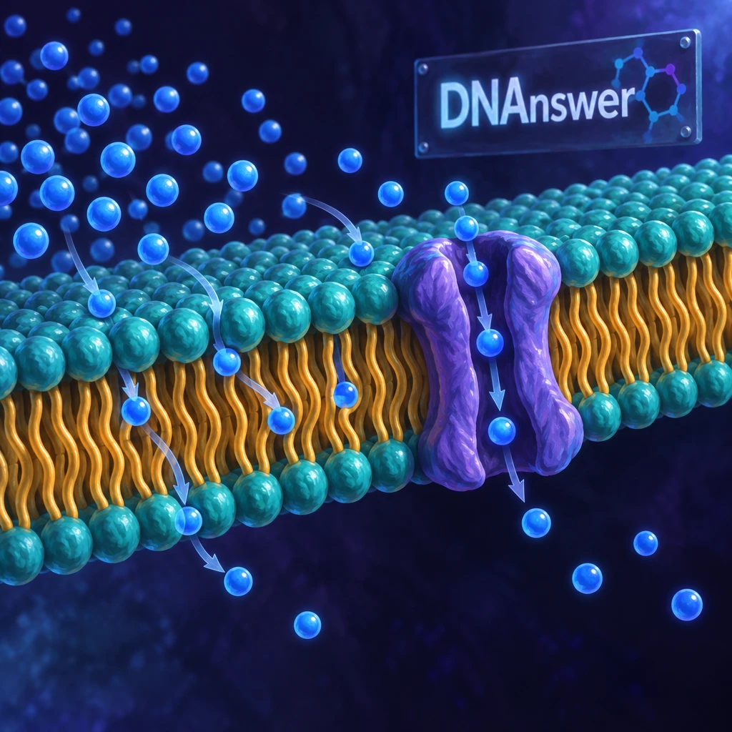
The real dividing line concerns energy, not proteins. Passive transport moves substances down a concentration difference, from more crowded to less crowded, and the cell spends no energy on it. That category includes two routes:
- Simple diffusion straight through the lipid bilayer, used by oxygen and carbon dioxide.
- Facilitated diffusion, where a channel or carrier protein provides a path, used by glucose and many ions.
Both routes stop once the concentrations even out. Facilitated diffusion has one extra limit: it can max out. A carrier protein handles one molecule at a time, so when every carrier is busy, adding more glucose outside won't speed things up. Think of a row of turnstiles at a stadium.
Water deserves its own mention. Osmosis means water moving across a selectively permeable membrane toward the side with more dissolved particles. Water trickles through the lipids slowly, but many cells add water channels called aquaporins to speed it up. Your kidneys rely heavily on them. Put a red blood cell in pure water and osmosis pulls so much water inside that the cell swells and bursts.
Where active transport begins
Active transport pushes substances against their concentration difference, uphill, so it needs energy. Cells usually pay with ATP, the molecule that works as the cell's energy currency.
The best-studied example is the sodium-potassium pump. Each cycle uses one ATP to push three sodium ions out and pull two potassium ions in. Your nerve cells depend on this gradient to fire signals, and the pump consumes a big share of the energy your brain burns at rest. Your stomach lining runs a different ATP-driven pump that loads protons into your stomach to make acid.
Some active transport works indirectly. A protein lets sodium flow back in downhill and uses that flow to drag a second molecule, like glucose, uphill alongside it. Your gut absorbs sugar this way. The cell still pays, just through the sodium gradient its pumps built earlier.
So when you sort transport, ask one question first: does the cell spend energy to make this happen? Proteins can show up on either side of the answer.
Myth 5: Cholesterol in your cells is just a problem
Cholesterol carries a bad reputation. Most people know it from blood tests and heart disease warnings, so finding it inside every animal cell membrane sounds alarming.
That worry has a real basis. High levels of certain cholesterol-carrying particles in your blood do raise the risk of clogged arteries. But that story concerns transport in the bloodstream. Inside the membrane, cholesterol does essential work, and your body makes much of it on purpose.
Cholesterol consists of four carbon rings fused together, a compact, stiff shape. It tucks in between phospholipids and acts as a fluidity buffer. On a warm day, it holds the phospholipids together and stops the membrane from getting too runny. When things cool, it wedges the tails apart so they can't pack into a stiff, gel-like layer. Either extreme would wreck the membrane: too fluid and it leaks, too stiff and proteins can't move or work.
The plasma membrane contains a lot more cholesterol than many of the membranes inside the cell. Plants use similar molecules called plant sterols, and bacteria mostly manage without. Your body temperature stays near 98.6°F, but your skin cells in a cold wind or your fingers in snow face much bigger swings, and cholesterol helps their membranes cope.
Keep the two stories apart. When you read "cholesterol" in a health headline, it almost always refers to blood levels. In cell biology, it refers to a structural ingredient you can't do without.
Myth 6: The membrane only controls what goes in and out
Ask most people what a membrane does and you'll hear "it lets things in and out." True, but that answer leaves out at least half the job.
The myth survives because transport makes the most visible, testable function. You can measure glucose entering a cell. Measuring a conversation between cells takes more work. For decades, many scientists themselves treated the lipid layer as a scaffold that held the important proteins in place.
Recognition and communication
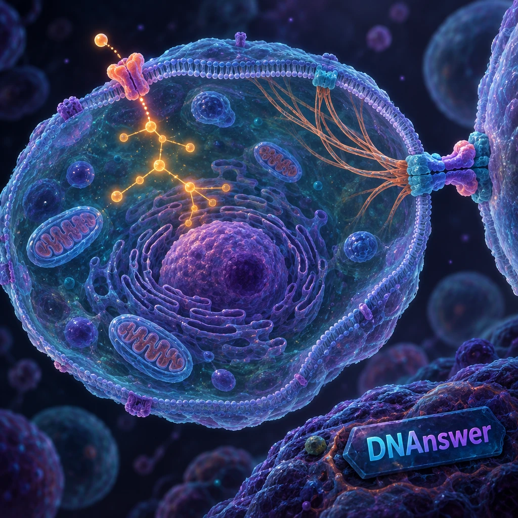
Sugar chains decorate the outer surface of the membrane, attached to proteins (forming glycoproteins) or to lipids (forming glycolipids). They appear only on the outside face, and they work like name tags. Your ABO blood type comes down to small differences in these sugar chains on your red blood cells. Your immune system reads such tags to tell your own cells from invaders.
Receptor proteins span the membrane and relay messages inward. Insulin, for example, never enters the cell to deliver its signal. It binds a receptor on the outside, and the receptor changes shape and triggers a response inside. We call this cell signaling.
The lipids matter more than anyone once thought. In April 2026, MIT chemists led by Gabriela Schlau-Cohen reported that membrane composition can change how a growth receptor called EGFR behaves. Normally about 15 percent of membrane lipids carry a negative charge. Membranes at 15 to 30 percent worked normally, but at 60 percent the receptor locked into an always-on state. That may help explain why some cancer cells divide uncontrollably.
Holding on and moving cargo
Anchor proteins let cells grip each other and hold onto internal support fibers called the cytoskeleton, a network of protein strands that gives the cell its shape. Your skin holds together under stretching partly because of these connections.
Membranes also move large cargo with bubbles. In endocytosis, the membrane folds inward around something outside, then pinches off into a sac inside the cell. Your white blood cells swallow bacteria this way. In exocytosis, a sac inside the cell fuses with the membrane and releases its contents outside. Nerve cells release chemical messengers like this at every signal, and your pancreas releases insulin the same way. This only works because the membrane flows, which brings you right back to Myth 1.
If you want to check how well these ideas stuck, you can try molecular biology quizzes in DNAnswer's free web app: https://dnanswer.app/quiz
Read the next diagram differently
The next time you see a membrane diagram, run three checks. Imagine every piece drifting. For any molecule crossing, ask about its size, charge and chemistry, then whether the cell spends energy. Look at the outer surface and name what each protein or sugar chain communicates. Those questions turn a static picture into a working system, and they apply to every cell you'll ever read about, from bacteria to neurons.
Frequently Asked Questions
Is the cell membrane the same as the plasma membrane?
Yes. Both names describe the same outer boundary of a cell. Biologists use "plasma membrane" to separate it from the many membranes inside a cell, such as the ones around the nucleus and mitochondria.
What does a cell membrane diagram show?
A typical diagram shows the two-layer phospholipid bilayer, proteins crossing or resting on it, cholesterol tucked between the lipids, and sugar chains on the outer face. Remember it shows one frozen moment. In reality, these parts move constantly.
What are the main components of the plasma membrane?
You'll find phospholipids, cholesterol, proteins and carbohydrates. In a typical human cell, proteins make up about half the mass, lipids about 40 percent and carbohydrates about 10 percent, though the mix varies between cell types.
Can anything cross the membrane without help?
Yes, small uncharged or fat-soluble molecules can. Oxygen, carbon dioxide and steroid hormones cross the lipid layer directly. Ions, glucose and larger molecules need channel proteins, carrier proteins, pumps or membrane sacs to get through.