Cytotoxic T Cells: How They Find and Kill Infected Cells
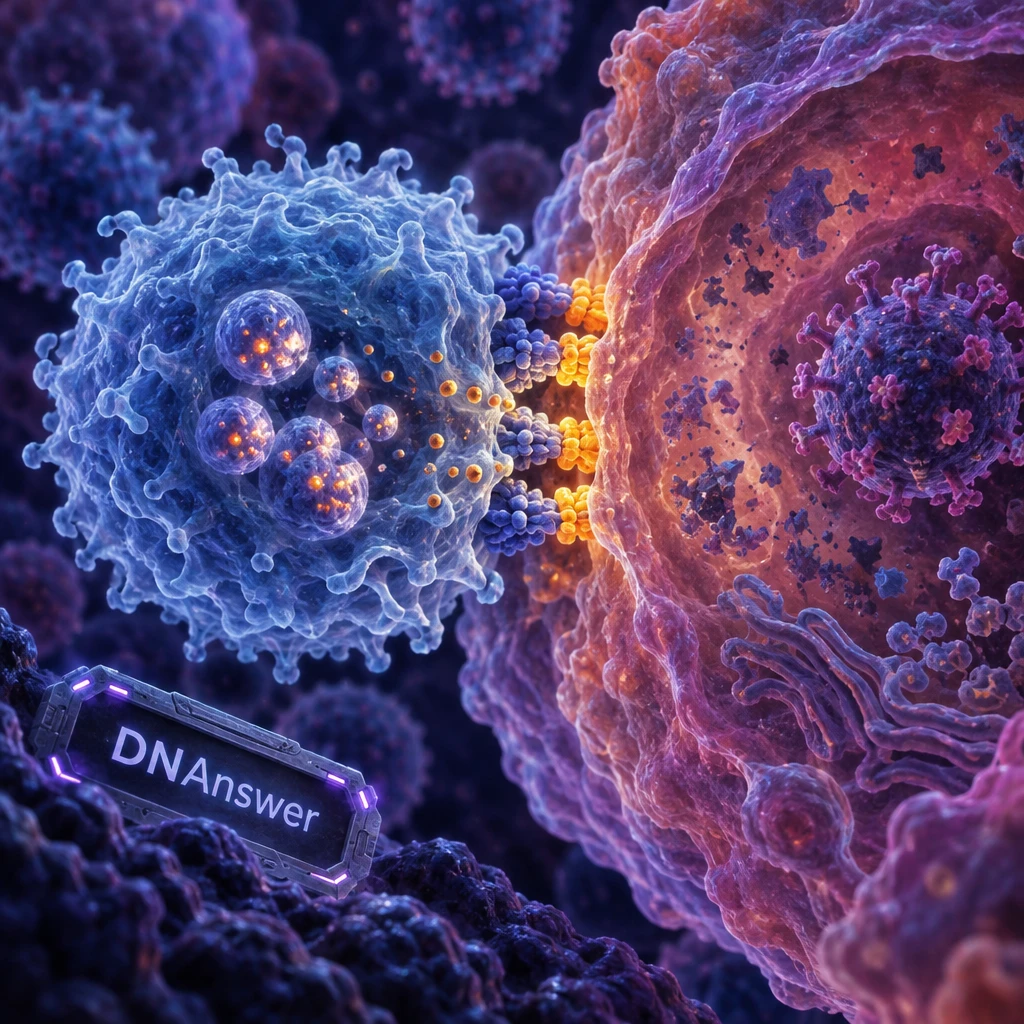
Do cytotoxic T cells kill infected cells the way soldiers kill enemies? Not quite. A cytotoxic T cell rarely destroys anything itself. It checks a cell's ID, then hands over the tools and signals that make the cell dismantle itself.
Cytotoxic T cells (also called CD8+ T cells or killer T cells) scan your cells for scraps of viral or abnormal protein shown on the surface. When a receptor matches a scrap, the T cell binds and releases perforin and granzymes, or uses a signal called Fas ligand. The infected cell then triggers apoptosis, a built-in self-destruct program.
Key Takeaways
- Killer T cells trigger the target's own self-destruct program, apoptosis.
- They see peptide fragments on MHC I, never whole viruses.
- Helper T cells coordinate; B cells make antibodies; CD8 cells kill.
- Naive killer T cells need activation by dendritic cells first.
- One killer T cell can kill several targets in sequence.
- Chronic infection and cancer can exhaust T cells, leaving them weak.
Myth 1: Killer T Cells Swallow and Digest Infected Cells
Picture a white blood cell wrapping around a virus-infected cell and digesting it. That image is common, and it has a real source. Phagocytes such as macrophages do exactly that: they engulf bacteria and debris and digest them. Textbook cartoons often draw all immune cells the same way.
Cytotoxic T cells work differently. They stay outside the target. After binding, they form a tight contact and deliver chemicals across it. Video microscopy of a T cell killing an infected cell shows the connection plainly: the T cell passes enzymes through it, and the target cell falls apart on its own schedule.
Assisted suicide, not murder
Apoptosis is programmed cell death, a tidy process cells already use to remove themselves during development or when damaged. The cell shrinks, chops up its own DNA and packages its remains into small bundles. Neighbors and cleanup cells then remove the bundles without any spill of inflammatory contents.
The T cell mostly supplies the trigger. Granzymes (enzymes that cut proteins) activate the cell's own caspases, a chain of protein-cutting enzymes that carries out the dismantling. The T cell starts the cascade. The target's machinery does the work.
What to do with this
Drop the "soldier" picture. Think of a T cell as an inspector who serves a notice. That shift explains why infected cells die cleanly and why viruses that block apoptosis can survive inside them.
Myth 2: T Cells Recognize Viruses Themselves
Antibodies grab whole viruses floating in your blood, so it seems natural that T cells do the same. Many people assume every adaptive immune cell spots the pathogen directly.
It doesn't work that way. Once a virus gets inside a cell, antibodies can't reach it. All viruses, and some bacteria, multiply inside the cell's interior. Only destroying the host cell removes them. So T cells need another way to see inside.
The cell's inside-out display
Every healthy cell constantly chops up some of its own proteins into short fragments called peptides. It loads these onto MHC class I molecules (proteins that act like small display trays) and carries them to the surface. A cell infected with a virus also chops up viral proteins, so viral peptides show up on the trays too.
A T cell never sees the virus. It sees a peptide on a tray. Its T cell receptor, or TCR, is a protein on the T cell's surface shaped to fit one specific peptide-and-tray combination. Your body makes millions of different TCRs, so some fit nearly any peptide.
The CD8 helper at the door
The CD8 protein on the T cell's surface grips the MHC tray itself and steadies the contact. That is why these cells are called CD8+ T cells. Almost every nucleated cell in your body carries MHC I, so killer T cells can check nearly all of them.
Takeaway: If a virus hides in your cells, T cells still find it, because the cell reports on itself.
Myth 3: Killer T Cells, Helper T Cells and Antibodies Do the Same Job
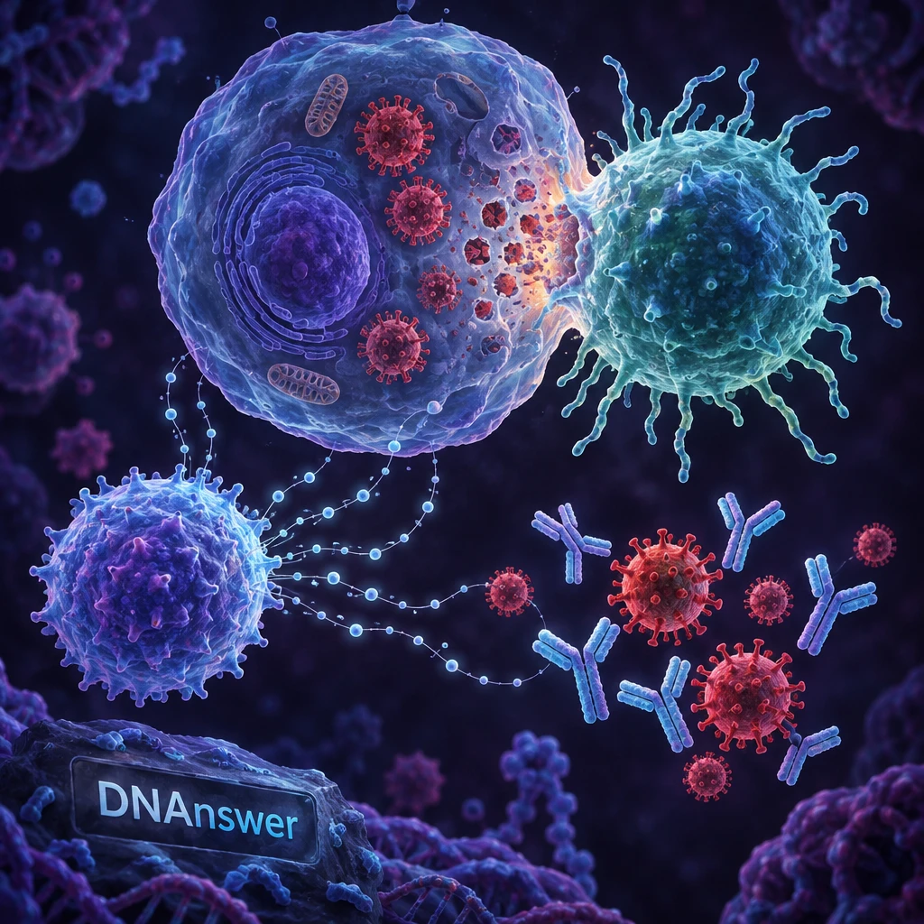
"T cells fight infection" is true, but it lumps together three different jobs. People mix them up for a fair reason: the names sound alike, and all of them belong to the same immune response.
Here is the split. CD8+ cytotoxic cells kill infected or abnormal cells. CD4+ helper T cells coordinate the response by releasing chemical messages called cytokines. B cells, a separate cell type, make antibodies. Antibodies are made by B cells, never by T cells.
The comparison, in plain terms:
- Marker on the surface: killer T cells carry CD8; helper T cells carry CD4.
- What they read: killer cells check MHC class I trays; helper cells check MHC class II trays, found on immune "display" cells.
- Main job: killers destroy infected cells; helpers coordinate other immune cells.
- Main tools: killers use perforin, granzymes and Fas ligand; helpers use cytokine signals.
- Where they act: killers act on any infected cell; helpers act mostly through immune cells.
Why the team matters
Helpers are not optional extras. They boost killer T cells and help B cells make better antibodies. HIV makes this vivid: it infects CD4+ helper T cells, and as they disappear, the whole response weakens. The AIDS lecture series that recorded killer T cell footage in 2007 made this link a core theme.
If you picture a hospital, killers are surgeons removing damaged tissue, helpers are the coordinators, and antibodies are the cleaning crew for the bloodstream.
Myth 4: Killer T Cells Attack Anything and Hurt Healthy Cells
If T cells are so deadly, why don't they destroy you? The worry is reasonable. Immune cells can harm healthy tissue, and autoimmune diseases such as type 1 diabetes show it happens.
Still, in normal life a killer T cell kills only after meeting a specific match. Removing infected cells without wrecking healthy tissue means the killing must be both powerful and accurate. Three safeguards make that work:
- Specific recognition. The TCR must fit one peptide on one MHC tray. A healthy cell showing ordinary self-peptides does not trigger it.
- Training. In the thymus, a small organ behind the breastbone, T cells that react strongly to your own proteins are mostly removed.
- A second signal. A naive T cell needs extra stimulation beyond the match alone before it can kill.
- Tight contact. The weapons go only into the one cell it is touching.
- Brakes. Receptors such as PD-1 and CTLA-4 slow T cells down when activity runs too high.
When safeguards fail
Failures are real but rare. Autoimmune disease happens when T cells that react to self slip through. Certain checkpoint cancer drugs release the brakes and can cause side effects such as inflammation in the gut or skin.
The practical point: "strong immune system" is not a goal. A balanced one is.
How Killer T Cells Find, Bind and Kill
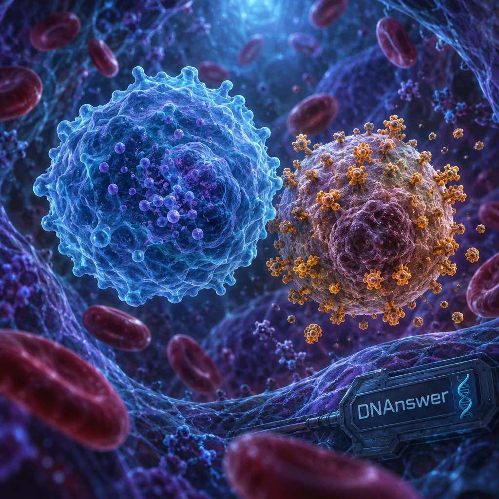
Here is the process from start to finish. Each step is a place where the system can be checked, and each one explains a different part of the "assisted suicide" idea.
- Scan. The T cell travels through blood and lymph, brushing against other cells and reading their MHC I trays.
- Match. If its TCR fits a peptide, it locks on with CD8's help.
- Seal. The two cells form an immunological synapse, a tight contact zone where receptors cluster and the cells seal off a small gap.
- Fire. The T cell moves granules (small packets) to the contact point and releases them into the gap.
- Punch. Perforin forms pores in the target's membrane.
- Trigger. Granzymes enter through the pores and start the caspase cascade.
- Detach. The T cell lets go and moves on.
Two ways to trigger apoptosis
The first route is the fast one. Perforin and granzymes act within minutes. The second is slower: the T cell shows Fas ligand on its own surface, which binds a Fas receptor on the target and sends a "die" signal inward. A 2011 review of how these two mechanisms work together noted that they account for most killing by these cells.
The T cell also releases interferon-gamma, a messenger that blocks viral copying and raises MHC I on nearby cells. That makes more infected cells easier to spot.
One cell, many kills
Yes, one killer T cell can kill more than one target. Because it detaches and moves on, it can kill several cells in sequence, and it often can do so within a short time. How many depends on how much ammunition it carries and how healthy it is.
How Killer T Cells Get Their License to Kill
A newly made killer T cell is naive: it has never met its peptide, and it can't kill yet. Many people assume your T cells are always armed. They are not, and that is a safety feature.
Priming by dendritic cells
Activation usually happens in a lymph node, a bean-sized filter station where immune cells gather. Dendritic cells are display specialists. They pick up viral material at a site of infection, travel to the node and show peptides to naive T cells. The T cell needs three things: a TCR match, a second "go" signal from the dendritic cell, and cytokines such as IL-2.
Once triggered, the T cell multiplies hard. One cell becomes a large clone, all with the same TCR, and these cells carry killing granules.
Where helpers come in
Helper CD4+ cells make the process stronger. They signal the dendritic cell to become a better activator and supply cytokines, including IL-2, that keep killer cells dividing. Without helpers, killer T cells can still start, but they often do worse over time.
If you wonder how a vaccine can train T cells, this is the answer: it gives dendritic cells something to show, so the response starts before a real infection.
When Killer T Cells Fail: Exhaustion and Evasion
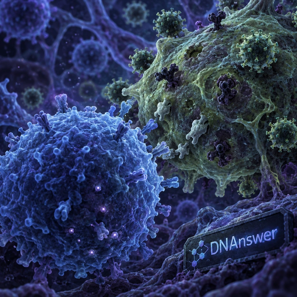
Constant exposure to a threat can wear killer T cells down. That sounds backwards, but it is how chronic infection and cancer often play out.
Exhaustion
Under long, steady stimulation, T cells enter exhaustion: they keep some activity but lose strength, make fewer cytokines and show more brake receptors such as PD-1, CTLA-4, TIM3 and LAG-3. One view is that exhaustion protects the body from damage caused by endless activation.
Immune checkpoint inhibitors have revolutionized the treatment of cancers such as melanoma, non-small cell lung cancer and renal cell carcinoma.
That finding, from a 2025 review in Signal Transduction and Targeted Therapy, shows why exhaustion matters. The U.S. Food and Drug Administration approved ipilimumab, a drug that blocks CTLA-4, for advanced melanoma. Such drugs release the brakes so T cells can attack tumors again.
Evasion
Viruses and tumors also hide. Some reduce the amount of MHC I on the cell surface, so the tray goes missing and the T cell sees nothing. Others block apoptosis or release signals that calm T cells. Another immune cell, the natural killer (NK) cell, helps here, since it attacks cells that lack MHC I.
Cancer cells are targets because mutations create odd proteins, and fragments of those appear on MHC I. That is why some cancer treatments aim at T cells.
What Cytotoxic T Cells Do and Don't Kill
They kill cells infected with viruses, cells with some bacteria living inside them, and cancer cells. They do not kill free-floating bacteria. Those are handled by antibodies, phagocytes and other defenses.
The distinction is about location. A bacterium in your bloodstream is outside cells, so antibodies and phagocytes reach it. A bacterium living inside a cell, such as the one that causes tuberculosis, is partly hidden, and T cells matter more.
If you want to test what you now know about T cells, MHC and apoptosis, DNAnswer has free immune system quizzes where you can earn XP and climb the leaderboard.
Conclusion
Next time you hear "the immune system attacks," ask which part. Killer T cells inspect, match and trigger a quiet self-destruct. Helpers coordinate. Antibodies handle what floats free. For decisions, that means a strong response depends on training (vaccines), balance (not overactive) and working brakes. If you read news about immunotherapy, look for whether it frees exhausted T cells or adds new ones.
Frequently Asked Questions
How do cytotoxic T cells find infected cells?
They scan MHC class I molecules on cell surfaces. If the T cell receptor matches a viral or abnormal peptide shown there, the T cell binds. Almost every nucleated cell displays these trays, so nearly any infected cell can be checked.
What do perforin and granzymes do?
Perforin punches holes in the target cell's membrane. Granzymes are enzymes that enter through those holes and activate caspases, which carry out apoptosis. Together they make the cell dismantle itself.
Do cytotoxic T cells kill bacteria?
Not directly. They kill host cells that harbor bacteria inside, such as cells infected with tuberculosis bacteria. Free bacteria in blood or tissue are cleared by antibodies and phagocytes.
What is the difference between cytotoxic T cells and helper T cells?
Cytotoxic T cells carry CD8 and kill infected or abnormal cells. Helper T cells carry CD4 and coordinate the response with cytokines. Neither makes antibodies; B cells do.