Function Cell Membrane: Structure and Transport
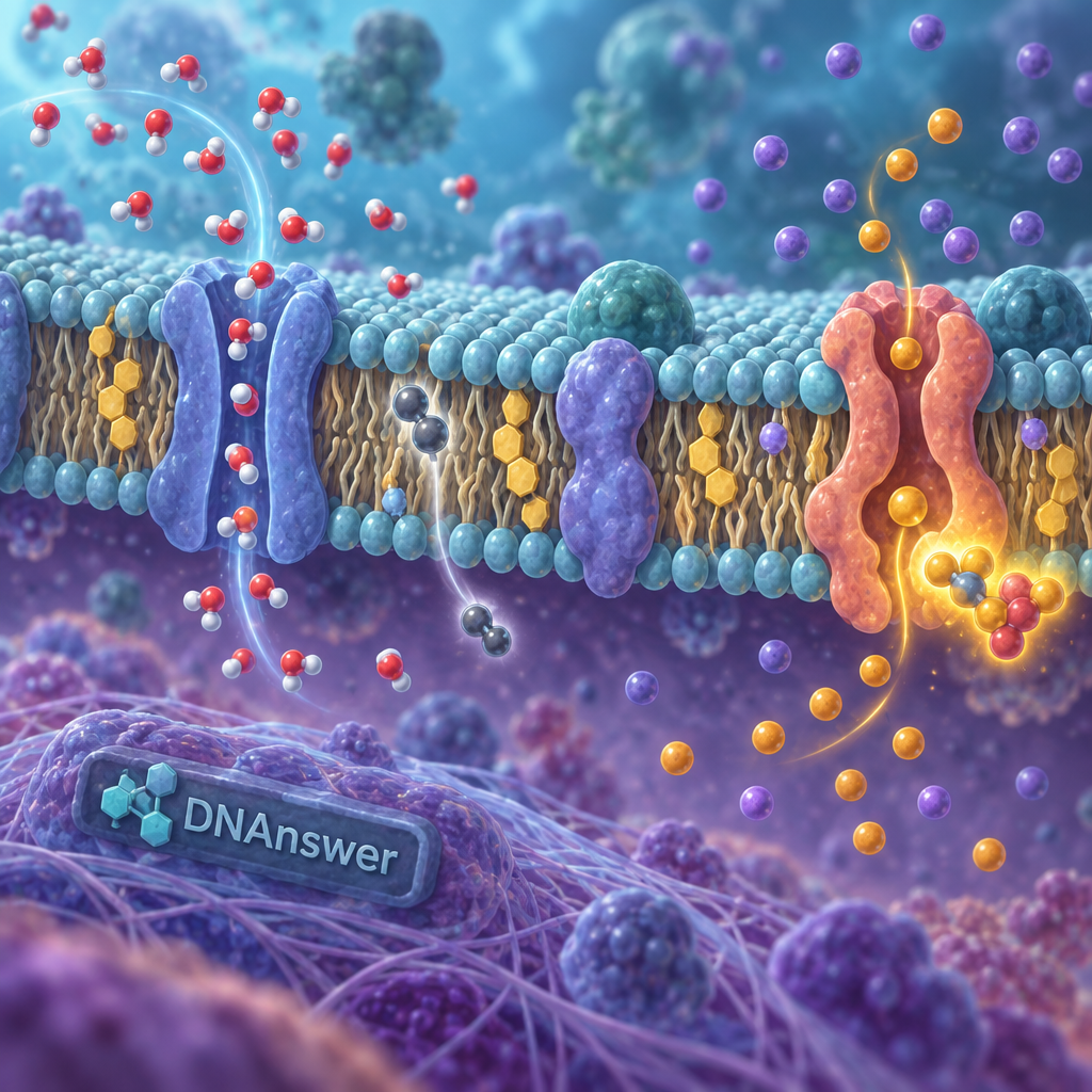
Your cell membrane is not a wall. It's closer to a bouncer at a club door, checking IDs and deciding who gets in, every second of your life. That's the real function of the cell membrane, and it's the opposite of what most diagrams in biology textbooks show you.
If you learned that the membrane is a static, plastic-bag-like boundary that just "keeps stuff in," you learned a shortcut that skips the most interesting part. The membrane is alive with motion, packed with proteins that make active decisions about what crosses it, and that decision-making is why cells can survive in salt water, stomach acid, or your bloodstream without falling apart.
This matters because the shortcuts stack up. Mixing up diffusion and osmosis, or confusing the membrane with the cell wall, leads to wrong answers on tests and a wrong mental model of how your own cells work.
Key Takeaways
- The membrane is a fluid mosaic, not a rigid shell — it flexes and shifts constantly.
- Only animal cells lack a cell wall; plants and bacteria have one plus a membrane.
- Selective permeability means the membrane blocks most molecules, not just some.
- Osmosis is a specific type of diffusion — water only, across a membrane.
- Membrane proteins run active transport, cell signaling, and even shape recognition.
- Cholesterol keeps the membrane from getting too stiff or too runny.
Myth: The Membrane Is a Rigid, Unchanging Wall
Picture a brick wall. Now picture a lava lamp. The cell membrane behaves far more like the second image, and that single correction unravels most of the confusion people carry about how cells work.
The belief that the membrane is fixed and wall-like comes honestly from how it's drawn. Most diagrams show a neat, static double row of molecules with a few proteins stuck in place like rivets. That image is technically accurate as a snapshot, but it freezes something that is never actually still.
The membrane is built from a phospholipid bilayer — two layers of fat-based molecules, each with a water-loving head and a water-hating tail, arranged tail-to-tail so the water-hating parts face inward, away from the watery environments on both sides of the cell. Individual phospholipids don't stay put. They slide sideways past their neighbors constantly, a behavior biologists literally call "fluid."
Why "Fluid Mosaic" Is the Accurate Name
In 1972, biologists S.J. Singer and Garth Nicolson proposed the fluid mosaic model, which describes the membrane as a shifting patchwork (mosaic) of lipids and proteins that move independently, like ice chunks floating and rotating on a pond. This model replaced older, more static views and remains the standard explanation taught today.
The fluid mosaic model describes the cell membrane as a two-dimensional fluid in which lipid and protein molecules diffuse more or less freely — Singer & Nicolson, Science, 1972.
What Actually Keeps It From Falling Apart
Cholesterol molecules wedge themselves between the phospholipids and act like a temperature regulator. When it's warm, cholesterol keeps the membrane from getting too loose and leaky. When it's cold, it stops the membrane from freezing solid. Without cholesterol, your cells would either dissolve or seize up depending on the weather.
The practical takeaway: if you're picturing the membrane as something that gets "damaged" like a torn bag, switch that image. Membrane damage usually means proteins misfolding or lipid composition shifting, not a rip in plastic. Cells actively repair and rebuild their membranes hour by hour.
Myth: The Cell Membrane and the Cell Wall Are the Same Thing
Ask ten people whether plant cells have a "membrane" or a "wall," and you'll get a coin-flip answer, because most people use the terms interchangeably. They shouldn't. This mix-up is one of the most common mistakes on any intro biology exam.
The confusion has a reasonable source: plant cells and bacteria have both structures, layered right next to each other, so diagrams often show them touching and looking like one thick boundary. Animal cells, on the other hand, only have the membrane, which makes the two structures easy to conflate if you only ever studied plant cells in class.
What the Cell Wall Actually Does
The cell wall is a rigid, non-living structure made of cellulose in plants (or peptidoglycan in most bacteria) that sits outside the membrane. Its job is structural: it resists pressure, holds the cell's shape, and stops the cell from bursting when water rushes in through osmosis. It doesn't select what enters or exits — it's more like scaffolding than a gate.
What the Cell Membrane Actually Does
The membrane, sitting just inside the wall, is the actual gatekeeper. It's flexible, alive, and involved in transport and communication. A plant cell without a wall (like a protoplast in a lab dish) can survive briefly, but it loses its shape immediately and becomes vulnerable to bursting, because the membrane alone can't handle pressure the way the wall can.
Animal cells never evolved a wall because they don't need rigid shape in the same way — your skin cells, muscle cells, and neurons rely on internal protein scaffolding (the cytoskeleton) instead. That's also why animal cells can change shape and squeeze through tight spaces, something a walled plant cell could never do.
If you remember one distinction: the wall protects structure, the membrane controls traffic. Mixing them up on a test, or in your mental model, means misunderstanding two completely different jobs.
Myth: Molecules Pass Through the Membrane Freely
Here's a fact that surprises most people: a glucose molecule sitting right outside your cell cannot just wander in. It needs a specific protein to escort it through, like a guest who needs a ticket scanned before entering an arena. This is the heart of selective permeability — the membrane's ability to let some substances through easily while blocking others almost completely.
The myth that everything passes through freely probably comes from diagrams showing tiny dots moving through the bilayer with arrows, which visually implies "anything goes." In reality, the bilayer itself is a fatty barrier, and only small, nonpolar (uncharged, fat-soluble) molecules like oxygen and carbon dioxide slip through the lipid layer directly.
Passive Transport: Moving Without Energy
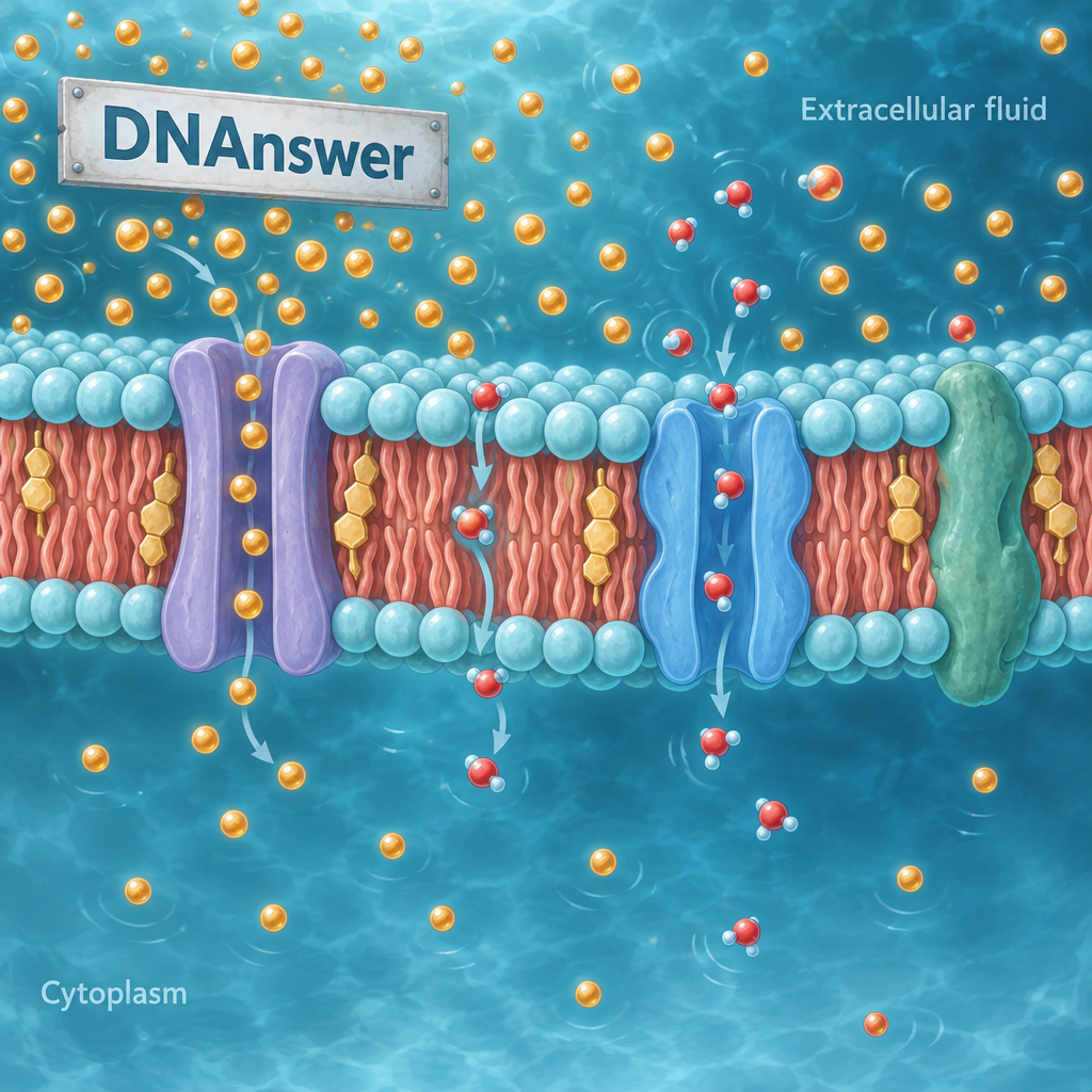
Passive transport happens when molecules move down their concentration gradient — from an area where they're crowded to an area where they're scarce — without the cell spending energy. This includes simple diffusion (small molecules crossing the lipid layer directly) and facilitated diffusion (larger or charged molecules moving through protein channels, like ion channels for sodium or potassium).
Active Transport: Moving With Energy
Active transport is different: it moves substances against their gradient, from low concentration to high, and that takes fuel. The sodium-potassium pump, found in nearly every animal cell, uses a molecule called ATP (the cell's energy currency) to push three sodium ions out for every two potassium ions it pulls in. This single pump uses roughly 20-40% of a resting neuron's energy budget, according to estimates from neuroscience research on metabolic cost.
Here's the sequence a molecule might face at the membrane:
- It arrives at the membrane surface and bumps into the lipid bilayer.
- If it's small and nonpolar (like oxygen), it slides straight through.
- If it's polar or charged (like glucose or sodium), it needs a channel or carrier protein.
- The protein checks the molecule's shape and size before allowing passage.
- If moving against the gradient, the protein needs ATP to power the move.
- If moving with the gradient, no energy is spent — the molecule just diffuses through.
The takeaway: assume nothing crosses without permission. Every substance that matters to your cell's survival, from sugar to sodium, is checked, scanned, and often pumped by a protein doing a specific job.
Myth: Osmosis and Diffusion Are the Same Process
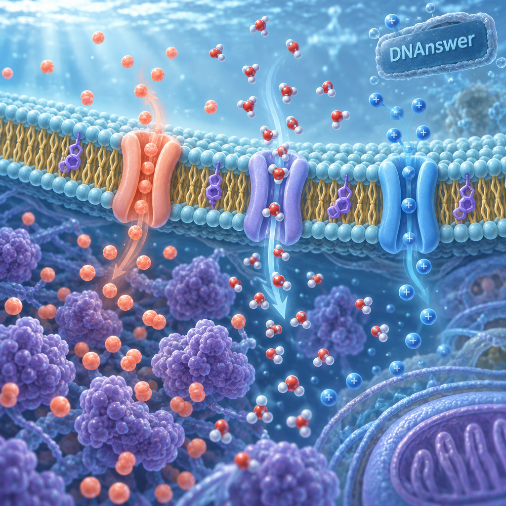
Diffusion and osmosis get taught back-to-back so often that they blur into one idea. They're related, but they're not interchangeable, and mixing them up leads to wrong predictions about what actually happens to a cell.
Diffusion is the general movement of any particle — gas, solute, ion — from high concentration to low concentration, anywhere, not just across membranes. Think of perfume spreading through a room. Osmosis is a specific, narrower case: it's the diffusion of water specifically, and specifically across a selectively permeable membrane.
Why the Distinction Actually Matters
If you put a red blood cell in pure water, water rushes in because of osmosis, and the cell can swell and burst. This happens because water moves toward the side with more dissolved solutes (salt, sugar, protein) to balance concentration on both sides. If you put the same cell in saltwater, water rushes out, and the cell shrivels. This is why IV fluids given at hospitals are carefully balanced (isotonic saline, at 0.9% sodium chloride) to match the salt concentration of blood — get that wrong, and you damage the patient's cells.
The Kernel of Truth Worth Keeping
Osmosis really is a form of diffusion, which is exactly why the two get merged. But calling all diffusion "osmosis" is like calling all vehicles "trucks." One is a subset of the other, and the distinction changes what you predict will happen to a cell in a given environment.
Next time you see a plant wilting despite regular watering, consider whether the soil has too much fertilizer salt — that pulls water out of root cells through osmosis, the opposite of what watering is supposed to do.
Myth: The Membrane's Only Job Is Keeping Things In and Out
This might be the most limiting myth of all, because it makes the membrane sound boring. In reality, the membrane is your cell's communication hub, not just its border patrol.
Embedded proteins called receptors stick out from the membrane surface and grab onto signaling molecules — hormones, neurotransmitters, growth factors — without letting the signal molecule itself enter the cell. The receptor changes shape when it binds its target, and that shape change triggers a cascade of reactions inside the cell. This is how insulin tells your muscle cells to absorb glucose, and how neurotransmitters trigger the next neuron to fire.
Endocytosis and Exocytosis: Bulk Transport
Sometimes a cell needs to move something too large for a channel protein, like a whole bacterium or a hormone-packed vesicle. That's where bulk transport comes in:
Endocytosis is when the membrane folds inward around a particle, wraps it in a pouch, and pulls it into the cell. Immune cells called macrophages use this constantly to engulf bacteria, in a specific version called phagocytosis. Exocytosis runs the reverse process: a vesicle inside the cell fuses with the membrane and dumps its contents outside, which is exactly how neurons release neurotransmitters into the gap between nerve cells.
Recognition: How Cells Know Friend From Foe
Membrane proteins also carry identification markers, similar to how your immune system tells your own cells apart from invaders. This is the basis for blood typing (A, B, AB, O markers on red blood cells) and for why organ transplants require matching donor and recipient tissue closely — mismatched surface markers trigger immune rejection.
Signaling and recognition, not just filtering, are core parts of the membrane's job description. Cut that function from your mental model, and you lose the explanation for how insulin works, how organ transplants fail, and how your immune system tells self from invader.
Fluid Mosaic in Practice: Why Membrane Shape Changes by Cell Type
Not every membrane looks or behaves the same way, and that variation is itself evidence against the "static wall" idea. A red blood cell's membrane is flexible enough to squeeze through capillaries a fraction of its own width. A neuron's membrane stretches across a signal-carrying extension (the axon) that can run several feet long in the human body.
Microvilli and Surface Area
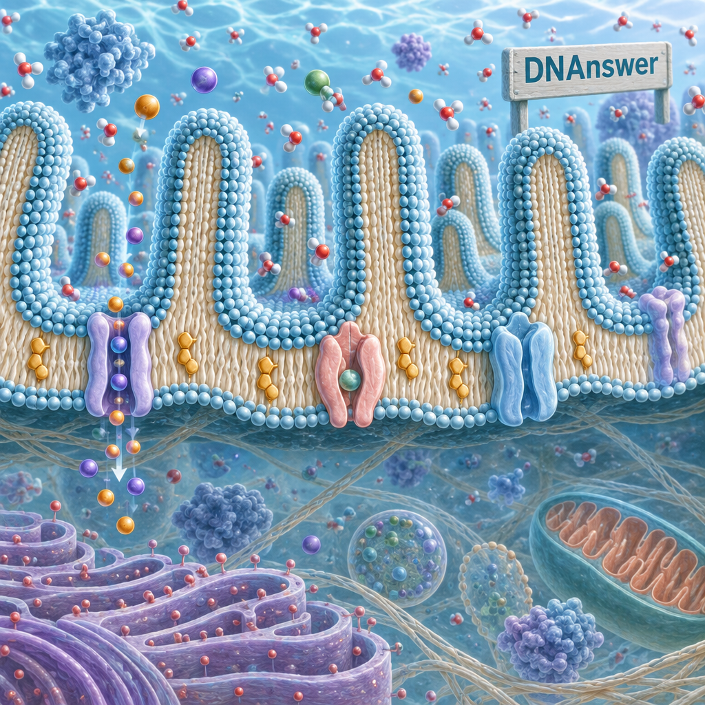
Intestinal cells fold their membrane into thousands of tiny finger-like projections called microvilli, which increase surface area dramatically, boosting nutrient absorption. Without this folding, digestion would take far longer, because there'd be less membrane surface available for transport proteins to work with.
Testing What You Know
If you want to check whether these ideas actually stuck, running through interactive quiz questions on membrane transport, like the ones built into the DNAnswer app, is a faster way to catch shaky spots than rereading a textbook page, because getting a question wrong forces you to pin down exactly which mechanism you mixed up.
Conclusion
Stop picturing your cell membrane as plastic wrap. Picture it as a border checkpoint staffed by thousands of specialized proteins, each trained for one job: pumping sodium, scanning for glucose, recognizing hormones, or engulfing invaders. When you're deciding how to explain osmosis, tonicity, or drug delivery to yourself, start from that checkpoint model — it predicts real outcomes, while the wall model only predicts wrong ones.
Frequently Asked Questions
What is the main function of the cell membrane?
Its main function is selective permeability — controlling exactly which substances enter or exit the cell using channel proteins, pumps, and receptors, rather than acting as a simple passive barrier that blocks or allows everything equally.
Do animal cells have a cell wall?
No. Animal cells have only a cell membrane; the rigid cell wall exists in plants (cellulose), fungi (chitin), and most bacteria (peptidoglycan), providing structural support the membrane alone can't offer.
Why does the sodium-potassium pump matter so much?
It maintains the electrical charge difference across neuron membranes needed for nerve signals, using ATP to move three sodium ions out for every two potassium ions in, against their natural gradients.
Can substances cross the membrane without any protein at all?
Yes, but only small, nonpolar molecules like oxygen, carbon dioxide, and some fat-soluble vitamins slip directly through the lipid bilayer; nearly everything else needs a channel, carrier, or pump protein.