Protein Structure: The Four Levels That Decide What a Protein Does
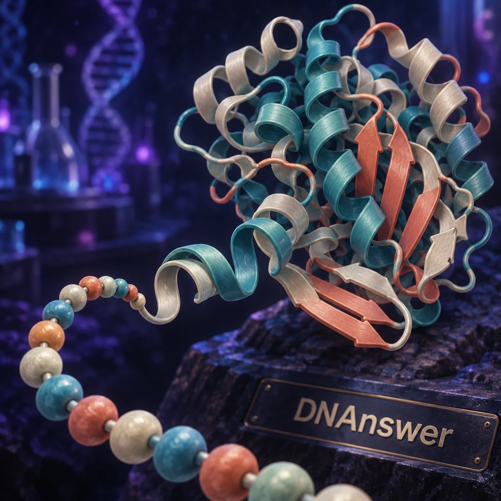
Does a protein really build itself in four neat steps, one level after another? No. The four levels are a teaching model, not a construction schedule. Primary structure is the amino acid sequence, secondary is local coils and folds, tertiary is the full 3D shape of one chain, and quaternary is several chains working together.
Your cells never build a protein "level by level." This guide shows where the tidy model works and where it breaks.
Key Takeaways
- Four levels describe a protein; cells don't build them as separate steps.
- Sequence largely sets the fold, but one swapped amino acid can wreck it.
- Many proteins are single chains with no quaternary structure.
- Denaturing unravels the fold, not the amino acid chain.
- AlphaFold predicts shapes well; it doesn't explain function on its own.
Myth 1: Function Comes From One Level of Structure
Plenty of quick summaries say the tertiary level "determines function." That is the most common shortcut, and it has a real basis. The 3D fold is the most direct factor, because it places the atoms that grab a target molecule or speed up a reaction.
But the fold doesn't appear from nowhere. The sequence dictates it, and local coils feed into it. In multi-chain proteins, the assembly adds another layer. Function emerges from all of them working together.
The Sickle Cell Example
Hemoglobin shows how one level reaches into the others. Swapping a single amino acid in the beta chain (glutamic acid replaced by valine) causes sickle cell disease. That is a primary-structure change. It alters how hemoglobin molecules stick to each other at the quaternary level, and red blood cells bend into sickle shapes. One letter in the sequence changed the whole outcome.
What to Do With This
Stop asking "which level matters?" Ask which level a problem starts at and where its effects show up. A mutation, a heat spike or a missing partner chain can each break function from a different entry point.
The Four Levels, Side by Side
Before correcting the rest of the myths, you need the baseline. Here is each level, what holds it together, and an example.
Primary structure is the order of amino acids (the small building blocks of proteins) in one chain. Peptide bonds, strong covalent links between neighboring amino acids, join them into a backbone. Insulin, for instance, has a specific sequence across two chains.
Secondary structure is a local pattern in the backbone. The two main patterns are the alpha helix (a coil) and the beta sheet (side-by-side strands). Hydrogen bonds between backbone atoms hold both. Silk fibroin is rich in beta sheets.
Tertiary structure is the whole folded shape of one chain. Side-chain interactions hold it: the hydrophobic effect, ionic bonds, hydrogen bonds and disulfide bonds. Insulin's disulfide bonds stabilize its fold.
Quaternary structure is the assembly of two or more chains, called subunits. The same kinds of interactions hold it together. Hemoglobin has four subunits.
Hydrogen bonds, ionic bonds and the hydrophobic effect are individually weak. Many of them together make a stable shape.
Myth 2: Every Protein Has All Four Levels
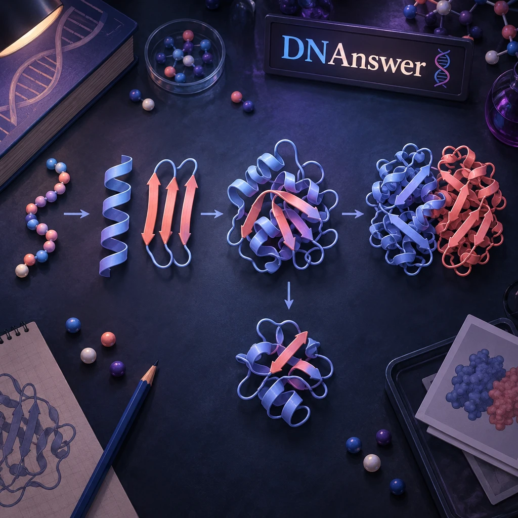
This belief comes straight from how the levels are taught. They are listed as a ladder, so it feels like every protein climbs all four rungs. And the best-known example, hemoglobin, does have all of them.
Reality is less tidy. Every protein has primary, secondary and tertiary structure, though the secondary part can be minor. Quaternary structure exists only when a protein is made of more than one chain. A protein built from a single chain stops at tertiary. It is complete and fully functional.
Plenty of proteins are single chains. Others are multi-chain: hemoglobin has two alpha chains (141 amino acids each) and two beta chains (146 each), and many enzymes work as dimers (two subunits) or tetramers (four). The hormone hCG has two chains.
Large proteins generally consist of several distinct protein domains, which are structural units that fold more or less independently of each other.
Domains matter here. A big single chain can contain several mini-folds, which is a different thing from several chains. Counting chains, not folds, tells you whether quaternary structure exists.
Takeaway: when you see "quaternary structure" on a quiz or in a paper, check the chain count first.
Myth 3: Proteins Build Themselves Level by Level
The ladder picture suggests a sequence: link amino acids, then make helices, then fold, then assemble. It's a fair way to teach, because each level really does depend on the one below.
In a cell, the steps overlap. A ribosome (the cell's protein-making machine) strings amino acids together, and the chain starts folding while it is still being made. Helices and the overall fold form together, not one after another.
Why the Sequence Still Rules
Test-tube experiments show how strong the sequence is. Scientists unfolded purified proteins with chemical solvents, then removed the solvent. Many proteins refolded spontaneously into their original shape. So the instructions for the fold sit in the sequence. A folded protein sits at its lowest free energy (its most stable arrangement).
Where Chaperones Come In
Cells are crowded, and unfinished chains can stick to each other. Chaperones are helper proteins that shield a chain while it folds and give it a second chance if it goes wrong. They don't dictate the shape. They keep the sequence's own instructions from being interrupted.
Takeaway: think of the levels as a description of the finished product, not a recipe.
Myth 4: A Protein Is a Rigid Sculpture
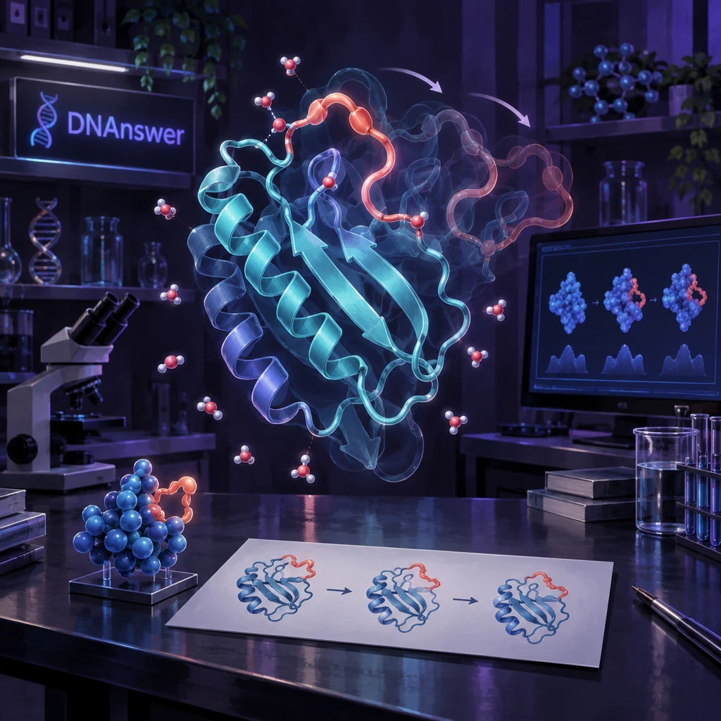
Ball-and-stick pictures in textbooks look like carved stone. That is a reasonable result of how structures are measured, since methods like X-ray crystallography capture a frozen snapshot.
Real proteins move. Enzymes flex as they bind a target. Some regions are intrinsically disordered, meaning they have no fixed shape at all and stay floppy by design. Those loose regions often act as flexible tethers or as switches that adopt a shape only when they meet a partner.
This breaks the "sequence makes one fixed fold" rule. For disordered regions, the sequence encodes flexibility, not a shape. The lesson: a static image is an average. When you see a clean helix in a diagram, ask what the protein does between snapshots.
Myth 5: Denaturation Breaks the Amino Acid Chain
Cooking an egg turns clear white to solid white. It looks like destruction, so it is natural to assume the protein got chopped up.
It didn't. Denaturation means unfolding. Heat, acid or solvents disrupt the weak interactions (hydrogen bonds, ionic bonds, hydrophobic packing) that hold secondary and tertiary structure. The strong peptide bonds of the backbone survive, so the primary structure stays intact. The chain becomes a flexible strand that has lost its shape and its function.
That is why a few cases reverse. When researchers removed the denaturing solvent, some proteins refolded. A cooked egg doesn't un-cook, because the unfolded chains tangle and clump together.
Misfolding Is the Cousin Problem
Misfolding means a chain folds into the wrong shape, often sticking to others. The A3D database applies predicted structures to rate aggregation tendencies across more than 20,500 human proteome entries. That kind of work links shape errors to disease.
Takeaway: fever and cooking both unfold proteins, but only digestion cuts the chain.
Myth 6: Sequence Alone Tells You the Shape, and AI Solved It
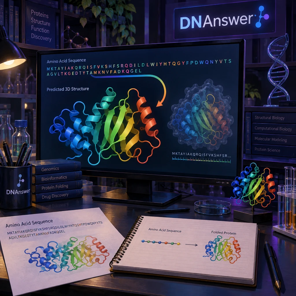
Both halves of this myth have a kernel of truth. The sequence does determine the fold in principle, as refolding experiments show. And AI has made huge progress. Predicting 3D structure from sequence was a goal for decades, and benchmarks such as CASP (Critical Assessment of Protein Structure Prediction) have run every two years since 1994.
Tools like AlphaFold and RoseTTAFold now predict most folds with high accuracy. In 2022, Oak Ridge National Laboratory ran full-proteome inference on 35,634 protein sequences using its Summit supercomputer.
What Prediction Does Not Give You
A predicted shape is one snapshot. It doesn't show motion, disordered regions or how a protein behaves with partners. Confidence also varies: researchers at the University of Oxford documented gaps in AlphaFold database confidence for proteins from Trypanosoma and Leishmania, parasites behind neglected tropical diseases.
Knowing the shape also doesn't tell you the job. Drug teams still combine predictions with simulations and lab tests, as one group did for the NLRP3 drug target.
How to Read a Prediction
- Check the confidence score before trusting a region.
- Treat low-confidence stretches as possibly disordered.
- Remember it shows one conformation.
- Look for whether partner chains were modeled.
- Confirm important claims with experiments.
If you want to test yourself on any of this, DNAnswer has free molecular biology quizzes that cover protein structure.
Two Protein Families That Break the Tidy Model
Fibrous and globular proteins show how different the same four levels can look.
Keratin (hair and nails) and collagen (skin, tendons) are fibrous proteins: long, rope-like and mostly insoluble in water. Their repetitive sequences build one dominant secondary pattern that gives strength.
Globular proteins, such as enzymes and hemoglobin, fold into compact balls that dissolve in water. Their hydrophobic side chains tuck inside and water-loving ones face outward.
Silk shows how sequence feeds structure: fibroin is rich in glycine, alanine and serine, which have small side chains, so beta sheets stack tightly.
Conclusion
Use the four levels as a map, not a manual. When you read about a mutation, a fever or an AI prediction, ask which level it touches and what else shifts. Then check what the model leaves out: flexible regions, single-chain proteins and moving parts. That habit will make most headlines about proteins easier to judge.
Frequently Asked Questions
What are the four levels of protein structure?
They are primary (amino acid sequence), secondary (helices and sheets), tertiary (the full 3D fold of one chain) and quaternary (several chains together). Not every protein has quaternary structure.
Which level of protein structure determines function?
The tertiary fold is the most direct factor, since it positions the active atoms. But sequence, local patterns and subunit assembly all contribute, so no single level works alone.
What bonds hold each level of protein structure together?
Peptide bonds link the primary chain. Hydrogen bonds hold secondary patterns. Tertiary structure uses the hydrophobic effect, ionic bonds, hydrogen bonds and disulfide bonds. Quaternary structure uses the same types of interactions between chains.
What happens when a protein is denatured?
It unfolds and loses its shape and function. The peptide bonds stay intact, so the sequence survives. Some proteins refold when the cause is removed, but cooked egg white doesn't.