Animal Cells: How Their Parts Work Together
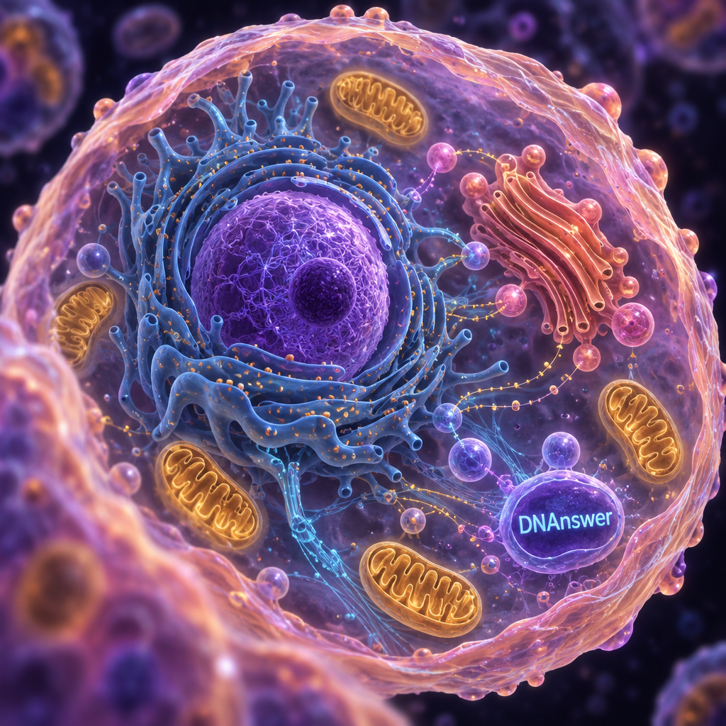
Is the nucleus really the boss of your animal cells? Not quite. It stores the instructions, but it can't build, fold, ship or power anything on its own. Animal cells run as a coordinated system: ribosomes build proteins, the endoplasmic reticulum and Golgi apparatus fold and package them, mitochondria supply energy, lysosomes recycle waste, and the membrane guards the border. Below, you'll bust five common myths and follow one protein from DNA to the cell surface.
Key Takeaways
- The nucleus stores instructions; other parts do the actual work.
- Organelles form a supply chain, not a memorization list.
- Mitochondria convert food into ATP; they don't create energy.
- Textbook diagrams are tidy; real cells are crowded and constantly moving.
- "13 parts" is a convention, not a fixed count.
Myth 1: The Nucleus Is the Boss, or the Brain
Picture a cell with its nucleus removed. It keeps running for a while, then fades. That fact alone should make you doubt the "boss" label.
The belief is understandable. The nucleus holds your DNA, and DNA carries the instructions for every protein. Many school materials call it the "brain" of the cell because it directs what happens inside. That shorthand has a kernel of truth: nothing gets built without those instructions.
But a brain senses, decides and commands. The nucleus does something closer to archiving. It keeps the master copy of your genes safe and hands out working copies on request. What gets copied depends on signals arriving from outside the nucleus: hormones landing on the cell surface, nutrient levels, damage alerts. The cell tells the nucleus what to read.
Why a library fits better than a boss
Think of the nucleus as a locked reference library. Your DNA is the one-of-a-kind book collection. Nobody removes the originals. Instead, the cell photocopies a single page, called messenger RNA (mRNA, a disposable copy of one gene), and sends it out the door.
The library also can't act on its own pages. It has no machines to read them, no loading dock and no power supply. Every one of those jobs belongs to something outside the nucleus.
What to do differently: when you see "nucleus = brain" on a flashcard, translate it to "nucleus = protected archive." The question to ask is always "who reads this copy next?" That question leads you straight into the rest of the cell.
Myth 2: Organelles Are Separate Parts on a List
You probably learned organelles (the cell's small working compartments) as a list: nucleus, ribosomes, Golgi, and so on. Lists are easy to test, so schools teach them that way. A list also makes it look like each part works alone.
That is the costly myth, because it makes cell biology feel like trivia. The parts only make sense as handoffs. A ribosome without a nucleus has nothing to read. A Golgi apparatus without an endoplasmic reticulum has nothing to package. Remove any link and the chain stops.
The fix is to follow a single job from start to finish. We'll do that in the next section with one protein, because a protein's trip touches almost every organelle you've memorized.
You'll also hear different counts: "five parts," "ten parts," "13 parts." Those numbers vary because authors group things differently. Some lists include the cytoskeleton, some skip it. Some count the cytoplasm (the jelly-like interior) as a part, others treat it as the space between parts. No official number exists, so don't memorize one.
One Protein's Journey: Five Handoffs From DNA to Membrane
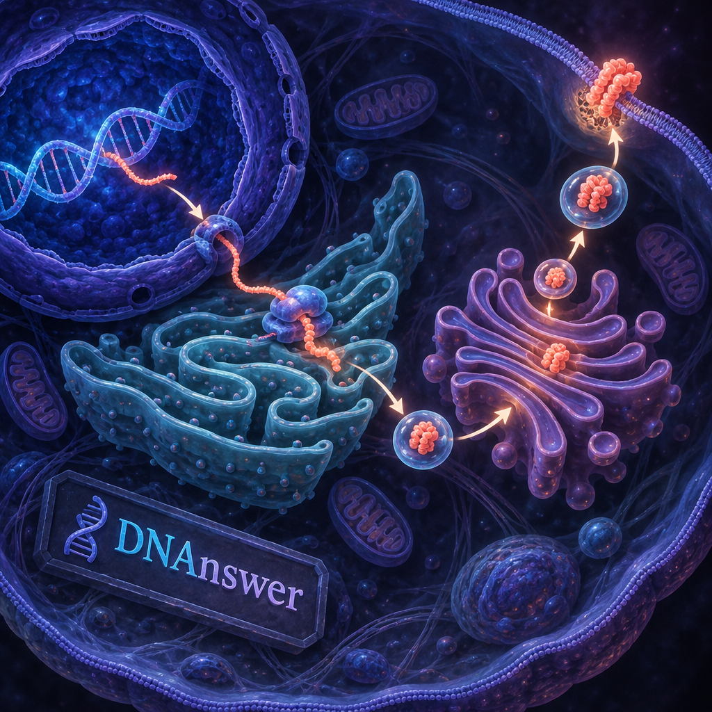
Say your cell needs to make a protein that sits in its outer membrane, such as a receptor that catches a hormone signal. This is the path it follows. Each step depends on the one before it.
- The nucleus copies the gene. A cell enzyme copies the gene's instructions from DNA into mRNA. The DNA never leaves.
- The mRNA exits and meets a ribosome. Ribosomes are tiny molecular machines that read mRNA and link amino acids (protein building blocks) into a chain. For membrane-bound proteins, this happens on the rough endoplasmic reticulum.
- The rough ER folds and checks the protein. The endoplasmic reticulum (ER) is a maze of folded membrane. The rough kind is studded with ribosomes. It twists the chain into its working shape and then pinches off a bubble called a vesicle (a small membrane-wrapped package) to carry it onward.
- The Golgi apparatus finishes and sorts it. The vesicle fuses with the Golgi apparatus, a stack of flat membrane sacs. Here the protein gets chemical tags, like sugar chains, that work as a shipping label.
- A second vesicle delivers it. The Golgi packs the protein into another vesicle, which merges with the cell membrane and places the protein where it belongs.
Notice what's missing from that list: any boss. Each step is a handoff, triggered by a physical event. The nucleus did its job in step one and then stayed out of it.
Not every protein takes this route
Free ribosomes floating in the cytoplasm make many proteins that stay inside the cell and do their work in the fluid there. Only proteins headed for the membrane, for export, or for certain organelles take the ER-to-Golgi route. This route is called the secretory pathway.
What powers the trip
Building and moving proteins costs energy. Much of that comes from ATP made by mitochondria, which brings us to the next myth.
Myth 3: Mitochondria "Make" Energy
"The powerhouse of the cell" may be the most repeated line in biology. It survives because it's catchy and partly right: mitochondria supply most of the usable energy in many animal cells.
But energy can't be created. Physics rules that out. Mitochondria are converters. They take molecules from food, mainly breakdown products of glucose (a sugar), and turn their stored chemical energy into ATP (adenosine triphosphate, the small molecule cells spend like cash). Muscle contraction and protein building both run on it.
The process is called cellular respiration, and it has three main stages. The first, glycolysis, happens in the cytoplasm and splits glucose into two smaller molecules. It needs no oxygen but nets only 2 ATP. The second, the citric acid cycle, runs inside the mitochondrion's inner space (the matrix). The third, oxidative phosphorylation, happens on the inner mitochondrial membrane.
Under ideal conditions, cellular respiration yields roughly 36 to 38 ATP per glucose molecule, but the real net yield is closer to 30 to 32. — Osmosis, cellular respiration overview
That gap matters. Cells lose some energy as heat and spend some on moving molecules around, so the textbook maximum is a ceiling, not a guarantee.
What to do differently: say "mitochondria convert," not "mitochondria make." You'll see why your body needs a constant supply of food and oxygen. Without raw material, the converters have nothing to convert.
Myth 4: The Textbook Diagram Shows What a Cell Looks Like
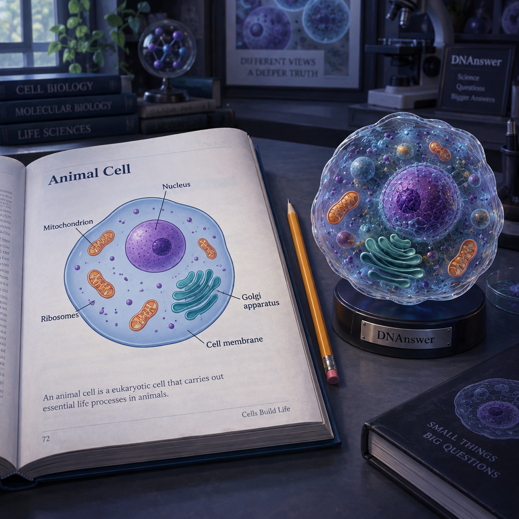
Open any biology book and you'll find an animal cell drawn as a neat oval. The nucleus sits in the middle, the organelles float in clean white space, and everything is color-coded.
That diagram exists for good reasons. You can't learn names and shapes from a blur, and a labeled cartoon gives you a map. The problem starts when you mistake the map for the territory.
Real cells are packed. The cytoplasm is thick with proteins, and organelles bump against each other all the time. Nothing sits still, either. Vesicles shuttle along tracks, mitochondria split, fuse and travel, and the ER stretches and reshapes itself.
The cytoskeleton: roads and scaffolding
The cytoskeleton is a network of protein fibers running through the cell. It does two jobs. It holds the cell's shape, since animal cells have no rigid wall. And it acts as a road system: motor proteins "walk" along the fibers, hauling vesicles to their destinations. That's how the vesicles in our protein journey get from the ER to the Golgi to the membrane instead of drifting randomly.
Shape follows job
There's no single "animal cell shape." A muscle cell is long and stretchy, a nerve cell has branches that can reach long distances, and a skin cell is flat. Specialization changes the contents too. A mature red blood cell has no nucleus, so it doesn't look like the cell in the diagram at all. Animal cells are usually between about 1 and 100 micrometers wide, so thousands could fit across a pencil tip.
What to do differently: treat any diagram as a legend, not a photo. When you study one, ask what moves and what it's stuck to.
Myth 5: The Membrane Is Just a Wall
Many people picture the cell membrane as a thin skin, like plastic wrap, that keeps the inside in. It does separate inside from outside, so the idea isn't wrong. It's just incomplete.
The membrane is a double layer of fat-like molecules called phospholipids. Their water-loving heads face the watery environments on both sides, and their water-repelling tails tuck inward. Cholesterol tucked between them adjusts how fluid or stiff the layer feels.
The real action comes from proteins embedded in it. Some are channels and pumps that move specific substances across. Some are receptors that catch signals, like the hormone receptor from our protein journey. Others anchor the cell to its neighbors. Because it lets some things through and blocks others, the membrane is called semi-permeable.
So the membrane isn't a wall. It's a border checkpoint with an inspection staff, and the staff is built from proteins that the rest of the cell assembled and shipped there.
That connects back to the supply chain. The membrane is both the final destination for many proteins and the thing that decides what the cell takes in next.
How Animal Cells Stay Together and Clean Up
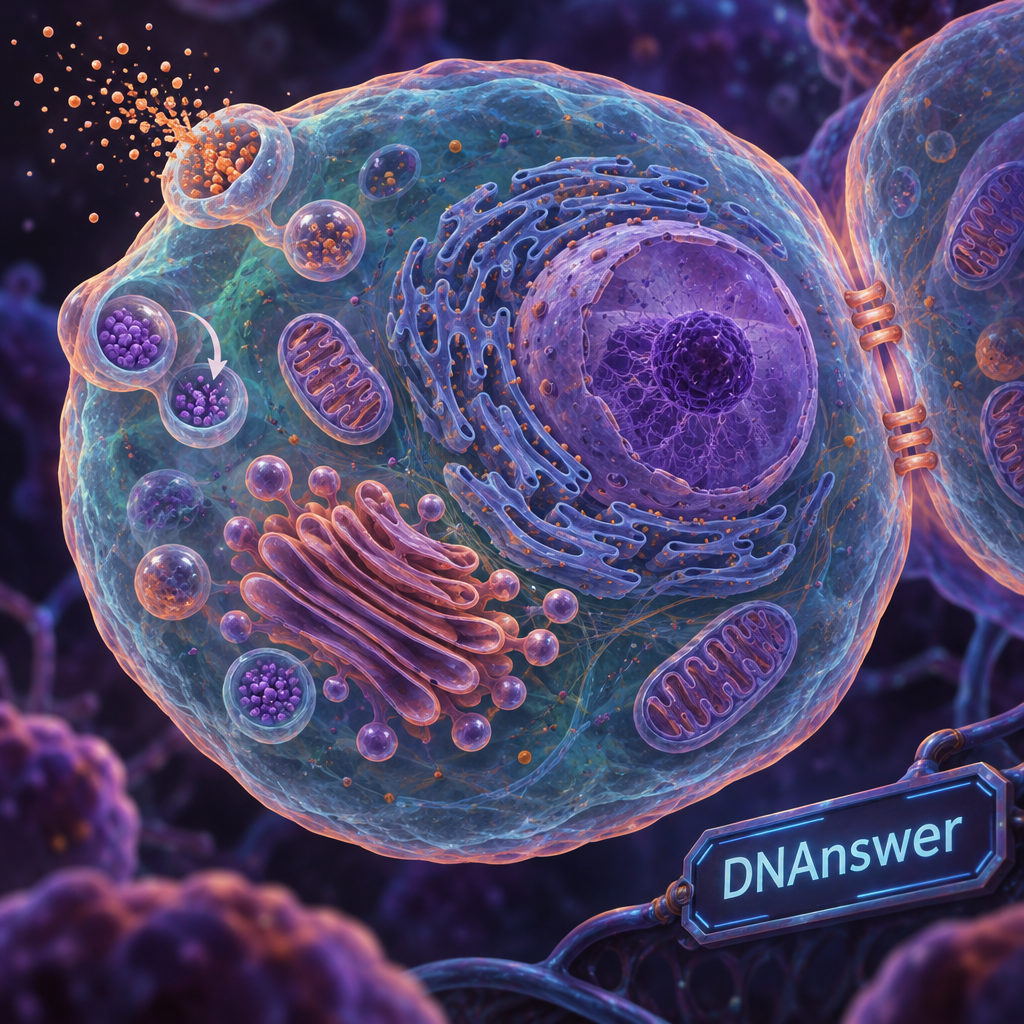
Two jobs finish the picture: recycling inside the cell and gluing cells to each other. Neither fits neatly into the protein story, but both keep it running.
Lysosomes: the recycling crew
Lysosomes are small sacs filled with digestive enzymes. They break down worn-out organelles, food particles and invaders, then release the pieces for reuse. If a cell is badly damaged, lysosomes can release their enzymes and dismantle the whole cell. Without them, waste would pile up like uncollected trash.
Don't assume other compartments share this job. Animal cells have vacuole-like sacs, but unlike plant cells they have no giant central one, and waste removal isn't their single role.
Cell junctions: how cells hold together
Animal cells don't have walls, so they use protein connections called junctions. Three main types do the work:
- Tight junctions seal gaps between cells so fluid can't leak through, as in your gut lining.
- Anchoring junctions link the cytoskeletons of neighboring cells, giving tissues strength where they stretch, like skin and heart muscle.
- Gap junctions form tiny tunnels that let small molecules pass directly from one cell to the next.
Together they turn billions of separate cells into tissues, and tissues into organs.
Animal vs. Plant Cells: What Actually Differs
Both are eukaryotic cells, meaning they have a true nucleus wrapped in a membrane. They share the nucleus, mitochondria, ER, Golgi and ribosomes. The differences are few, but they change how each cell lives.
Plant cells have a rigid cell wall, a large central vacuole full of water that keeps the cell firm, and chloroplasts, which capture sunlight for photosynthesis. Animal cells lack all three. Many animal cells also have a centrosome, which helps organize the separation of DNA during cell division.
The missing wall is a big deal. Without it, animal cells can take many shapes, which allows the huge range of cell types in your body, from neurons to muscle. Plant cells are more regular because the wall locks their form.
If you'd like to test whether the handoffs have stuck, the free cell biology quizzes on DNAnswer let you practice and track your progress.
Conclusion
Next time you see a cell diagram, trace a path instead of reading labels. Pick one protein and ask who reads its instructions, who folds it, who tags it, who carries it and who pays the energy bill. If you can narrate that chain without checking notes, you understand the cell better than a list ever taught you.
Frequently Asked Questions
How do the parts of an animal cell work together?
They work as a supply chain. The nucleus stores DNA, ribosomes build proteins, the ER and Golgi fold and ship them, mitochondria supply ATP, and the cytoskeleton moves cargo. Lysosomes recycle waste, and the membrane controls what crosses the border.
How does a protein travel from the nucleus to the cell membrane?
An mRNA copy of the gene leaves the nucleus and reaches a ribosome on the rough ER. The ER folds the protein, a vesicle carries it to the Golgi, and the Golgi sorts it and ships it to the membrane in another vesicle.
Is the nucleus really the brain of the cell?
No. The nucleus is a protected archive of DNA. It copies genes on request, but the cell's signals decide which genes get read, and other organelles do all the building, shipping and powering.
What is the difference between animal and plant cells?
Plant cells have a cell wall, a large central vacuole and chloroplasts. Animal cells have none of these, so they take more shapes. Many animal cells also have a centrosome, which helps organize cell division.